A synapse is the junction between the axon terminal of one neuron and the dendrite of the next, where signals cross from one cell to another. Most synapses are chemical: the sending neuron releases neurotransmitters such as dopamine, serotonin, or norepinephrine, which bind to receptors on the receiving neuron. Some synapses are electrical, with neurons connected directly through gap junctions.
Our body has a huge number of neurons. How many, you ask? Well, the human brain alone boasts around 86 billion neurons. That’s a very big number! These neurons are a part of our nervous system, which plays a very important role in our lives. It receives signals, processes them, and then decides and executes the appropriate outcome. As any system does, the nervous system has its own functional unit – a neuron. These neurons run throughout our body, intercepting signals and passing them to the brain or spinal cord. These neurons send information in a special manner, through synapses!
Structure Of A Neuron
To understand what synapses are and how they function a quick description of the neuron is necessary.
Neurons can seem pretty complicated, but their basic structure is quite simple. There are three broad parts – a cell body, a long (sometimes very long) axon and axon terminals. The cell body that contains the nucleus and extending from it are long dendrites that look like branches from a tree. In neuron communication, it is these dendrites that receive the signals. On the other end is the axon, which is a long, thin structure like the tail of the neuron. It is responsible for conducting or passing on the signals.
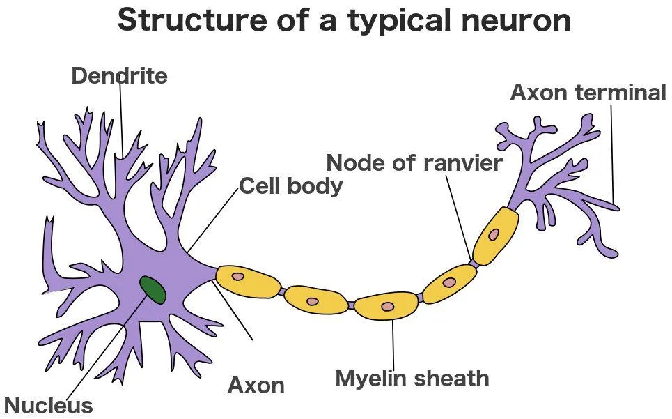
From the cell body extends the axon. It is a long tubular extension of the cell, and the axons of some neurons are wrapped in segments of a fatty insulating layer called the myelin sheath. These segments aren’t continuous; small gaps between them, known as the nodes of Ranvier, allow the signal to jump from node to node. The myelin sheath helps in the faster conduction of signals. A neuron will have many dendrites, but only a single axon.
The axon, near the end, branches off and forms axon terminals. At these terminals, a tiny bulb-like structure is present, which facilitates the passage of information between 2 neurons. More specifically, it facilitates the passage between the axon of one neuron and the dendrites of the next neuron. And this leads us to what a synapse is.
Synapse And Neurotransmitters
The juncture between the axon terminals of one neuron and the dendrites of another is known as a synapse. Synapses can be either electrical or chemical.
In a chemical synapse, the axon terminal sends information by releasing chemicals called neurotransmitters, which are then sensed by the dendrite of the other neuron. In electrical synapses, neurons are physically connected to each other through channels called ‘gap junctions’, causing direct electrical transmission.
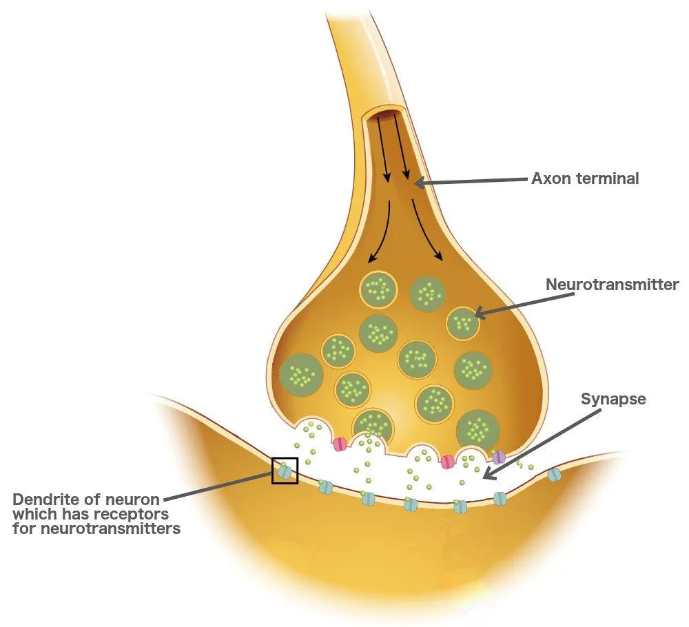
Neurotransmitters are chemicals produced in the cell body, transported to the axon terminal where they are stored. When an electrical signal reaches the end of a neuron, it activates the release of these molecules. The neurotransmitters then cross the gap to the dendrites of the next neuron, which have neurotransmitter receptors. The neurotransmitter receptors attach to the neurotransmitter, sensing the signal. These convert the chemical signal to an electrical impulse. If enough neurotransmitters have bound to the receptor, generating enough electrical potential, the signal will be conducted through the neuron until it reaches the axon terminal, where the same process is repeated. Different neurons orchestrate the release of different types of neurotransmitters, which, in turn, elicit an electrical impulse in a neuron with which it forms a synapse.
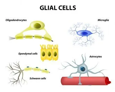
Some examples of neurotransmitters are norepinephrine, dopamine, and serotonin. Once these transmitters have evoked the desired response in the next neuron, they are either taken back up by the axon terminal that released them, mopped up by glial cells like astrocytes, or broken down by enzymes in the synaptic cleft. This is a necessary step, as it prevents the same signal from being sent over and over again, even when its stimulus ceases to exist. For example, to contract a muscle, the appropriate motor neurons release the neurotransmitter acetylcholine. If acetylcholine is not degraded (or removed from the site), then the muscle would constantly contract!
Given that the signal being conducted by the neurons is in the form of an electrical impulse, the synapse helps modulate signal being transmitted by using neurotransmitters. This is not possible in electrical synapses, where there is direct and instantaneous signal transmission from one cell to another.
Some neurons, though, are connected through electrical synapses rather than the chemical ones discussed above. This isn’t as common as the neurotransmitter mode of communication, but they exist. The axon terminal of these neurons is connected by gap junctions. These gap junctions are small channels that connect the two neurons and allow ions to freely pass through them. This makes passing the signal faster than the chemical synapse.
Synapses are present throughout our body. Our neurons fire signals at astonishing rates, and thus our synapses have to be very efficient at conducting the signals. Even though we perform multiple tasks at a time, our neurons and synapses never mix up the signals, thus ensuring the smooth functioning of the body.
How Does A Synapse Work, Step By Step?
So far we have seen the cast of characters, but how does a single message actually make the jump? At a chemical synapse, the whole event unfolds in roughly a thousandth of a second, and it always runs in one direction: from the axon terminal of the sending (presynaptic) neuron to the dendrite of the receiving (postsynaptic) one. Let’s walk through it.

First, an electrical impulse known as an action potential races down the axon and arrives at the terminal. That arrival flips open voltage-gated calcium channels, and calcium ions (Ca2+) rush into the terminal. This calcium surge is the trigger: it causes the tiny membrane-bound sacs called synaptic vesicles, each packed with neurotransmitter, to fuse with the cell membrane and dump their contents into the gap.
That gap is the synaptic cleft, and it is astonishingly narrow, only about 20 to 40 nanometres (a nanometre is a millionth of a millimetre) across. The neurotransmitter molecules diffuse across this sliver of space and lock onto matching receptors on the postsynaptic membrane, the way a key fits a lock. Binding opens ion channels on the receiving neuron, converting the chemical message back into an electrical one. In effect, a synapse turns an electrical signal into a chemical signal and then back into an electrical signal again.
Finally, the message has to be switched off so the next one can get through cleanly. The leftover neurotransmitter is cleared in one of three ways: it is pumped back into the sending terminal (reuptake), broken apart by enzymes in the cleft, or soaked up by neighbouring glial cells. Only then is the synapse reset and ready to fire again.
What Does A Synapse Do? Excitatory And Inhibitory Signals
It would be easy to picture a synapse as a simple on/off switch, but that sells it short. Its real job is to help decide whether a message gets passed along at all, and synapses come in two flavours that pull in opposite directions.
An excitatory synapse nudges the receiving neuron closer to firing. When the neurotransmitter binds, it produces a small positive blip in voltage called an excitatory postsynaptic potential (EPSP), making an action potential more likely. Glutamate is the brain’s main excitatory messenger. An inhibitory synapse does the reverse: it produces an inhibitory postsynaptic potential (IPSP) that makes the receiving neuron less likely to fire. GABA (gamma-aminobutyric acid) is the chief inhibitory neurotransmitter.
Here is the clever part. A single EPSP is usually far too weak to set off an action potential on its own. Most neurons in the brain are contacted by thousands of synapses at once, and the cell adds up all of these excitatory and inhibitory inputs, a process called summation. The neuron tallies the positives and negatives arriving at the same moment (spatial summation) and in quick succession (temporal summation). Only if the running total crosses a firing threshold does the neuron send its own signal onward. So a synapse is less an on/off switch and more a vote, and every neuron is constantly counting the ballots before it decides to fire.
How Many Synapses Are In The Brain, And How Do They Store Memories?
If the brain’s 86 billion neurons are impressive, the number of synapses between them is staggering. Each neuron can form thousands of connections, and the adult human brain is estimated to hold on the order of a hundred trillion synapses. That dense web is exactly what lets the brain do something a simple wiring diagram cannot: it can change.
The connections are not fixed. A synapse can grow stronger or weaker depending on how often it is used, a property neuroscientists call synaptic plasticity, and it is widely regarded as the cellular basis of learning and memory. The catchphrase is “neurons that fire together, wire together”: when two connected neurons are repeatedly active at the same time, the synapse between them is reinforced.
Two opposing processes do the tuning. In long-term potentiation (LTP), repeated activation makes a synapse larger and more effective, so the same input produces a bigger response. In long-term depression (LTD), low-frequency activity weakens the connection instead. These changes can last anywhere from hours to a lifetime, depending on how often they are reinforced. So when you memorise a phone number or learn to ride a bike, you are not so much adding new wires as quietly adjusting the strengths of synapses you already have. Your memories live, in a very real sense, in the changing pattern of those connections.
References (click to expand)
- Action potentials and synapses - Queensland Brain Institute. The Queensland Brain Institute
- Neurons, Synapses, Action Potentials, and Neurotransmission. Illinois State University
- Harris, K. P., & Littleton, J. T. (2015, October 1). Transmission, Development, and Plasticity of Synapses. Genetics. Oxford University Press (OUP).
- NEURONS - www.indiana.edu:80
- Neuroscience For Kids - questions/answers. The University of Washington
- Physiology, Neurotransmitters. StatPearls. NCBI Bookshelf.
- Summation of Synaptic Potentials. Neuroscience (2nd edition). NCBI Bookshelf.
- Kennedy, M. B. (2016). Synaptic Signaling in Learning and Memory. Cold Spring Harbor Perspectives in Biology.
- Mapping the Brain and Its Functions. Advancing Neuroscience in the Decade of the Brain. NCBI Bookshelf.