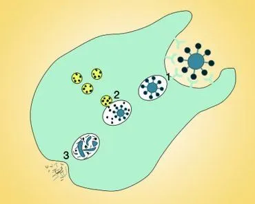
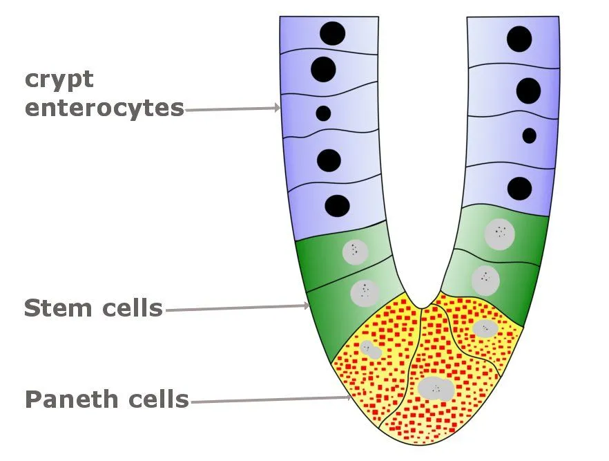
Paneth cells sit at the base of the small intestine’s crypts, alongside the stem cells that constantly renew the gut lining. When triggered by bacteria, they release a cocktail of antimicrobial molecules (α-defensins HD5 and HD6 in humans, lysozyme, and phospholipase A2) that punch holes in microbial membranes and help shape which microbes get to live in the gut.
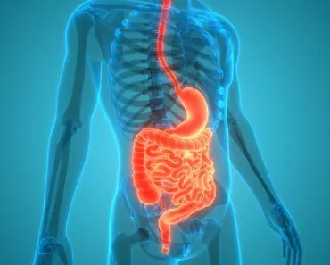
The food we consume goes through a long journey before it gets digested or excreted from the body. However, there are only a few organs that are directly involved in the process. One is the stomach, of course, and the other crucial part of the entire digestive process is the small intestine.
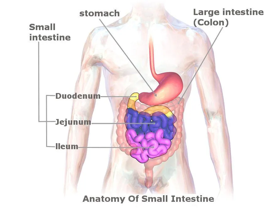
Most of us, who have little or no knowledge of the process of digestion, would simply assume that the stomach is the place where most of the digestive process occurs. However, that’s not really true. It’s the small intestine where 90% of the digestion and absorption of food happens in our body.
The small intestine is responsible for absorbing nutrients and minerals from food. Once the nutrients are broken down, they are absorbed by the inner walls of the small intestine and can flow into the bloodstream. The nutrients become so small there that they can pass across the epithelial cells of the gastrointestinal tract.
Paneth Cells
Paneth cells are one of the main cell types of the epithelium, which provide host defense against microbes in the small intestine. When these Paneth cells are exposed to bacteria, they secrete a number of antimicrobial molecules that help to maintain the gastrointestinal barrier.
For starters, the gastrointestinal barrier is a barrier formed by the gastrointestinal mucosa between the body and the lumenal environment. This environment is laden with potentially hostile toxins and microorganisms, in addition to nutrients that are beneficial to us. Thus, the gastrointestinal barrier helps keep that environment from interacting with the body (Source).
The main cell types of the epithelium (one of the types of animal tissue, i.e., epithelial tissues line the outer surfaces of blood vessels and organs, and inner surfaces of cavities in many internal organs) of the small intestine include enterocytes, goblet cells, enteroendocrine cells, tuft cells, and Paneth cells. All five are descended from Lgr5+ intestinal stem cells that live at the base of the crypts. A sixth specialized type, the microfold (M) cell, sits over Peyer’s patches and samples antigens for the immune system.
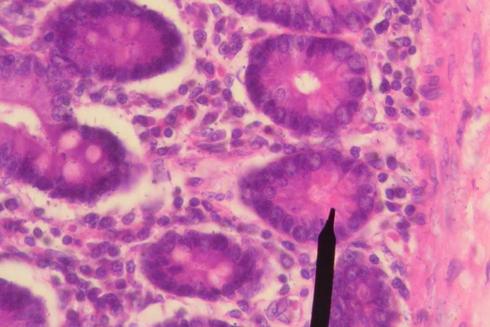
Of those types, Paneth cells are the easiest to spot under the microscope. They’re pyramid-shaped, packed with bright eosinophilic (pink-staining) granules, and they nestle right at the bottom of the crypt, between five and twelve to a crypt, with the densest populations in the ileum (the last stretch of the small intestine).
Paneth Cell Function
The main defensive molecules secreted by Paneth cells are alpha-defensins. In humans, the two stars of the show are HD5 (gene DEFA5) and HD6 (gene DEFA6). Most of the early Paneth-cell biology was actually worked out in mice, where the orthologous family is called cryptdins (short for “crypt defensins”), with about 17 isoforms. Cryptdins and HDs do the same job, but the names aren’t interchangeable: cryptdins are a mouse-specific term, while HD5 and HD6 are what you’ll find in your own gut.
The two HDs even split the work in interesting ways. HD5 is the killer: it punches directly into bacterial membranes and disrupts them. HD6 is the trapper: it self-assembles into thin protein nanonets in the gut lumen that physically snare bacteria before they can touch the epithelium (a clever little fishing-net trick, reported by Hooper and colleagues at UT Southwestern).
The killing mechanism comes down to chemistry. Bacterial membranes carry an unusually high concentration of negatively-charged phospholipids on their outer face, while our own cell membranes mostly hide their charged lipids on the inner leaflet. Defensins are small, positively-charged peptides, so they’re drawn electrostatically to bacterial surfaces, insert themselves into the bilayer, and form pores. The bacterium’s contents leak out, and our cells are largely spared.
Paneth Cells’ Role In The Small Intestine
When Paneth cells are exposed to bacteria (both gram-negative and gram-positive types) or to bacterial fragments such as lipopolysaccharide and muramyl dipeptide, they are stimulated to dump the contents of their granules into the crypt lumen. The granules contain defensins, lysozyme (an enzyme that chews up bacterial cell walls), and secretory phospholipase A2 (which cuts up the lipids in microbial membranes). Together, the three are a one-two-three punch against invaders.
Paneth cells boast a formidable arsenal thanks to these secretory molecules, which help them fight bacteria, fungi, and a surprisingly wide range of viruses. The virus part is worth dwelling on: alpha-defensins are unusual because, unlike most antimicrobial peptides, they’re effective even against non-enveloped viruses. HD5 in particular has been shown in the lab to neutralize adenoviruses, polyomaviruses (like BK and JC virus), and human papillomaviruses, all of which lack the lipid envelope that most antiviral peptides rely on.
In addition to offering protection against a wide variety of disruptive agents, Paneth cells are also capable of responding to ‘intestinal insults’ by dedifferentiating. After radiation injury, chemotherapy, or severe inflammation, post-mitotic Paneth cells can essentially turn back the developmental clock, re-enter the cell cycle, and act as backup stem cells that help repopulate the damaged crypt. The reversion is driven by reactivation of Notch signaling and SCF/c-Kit signals between the Paneth cell and its neighbors.
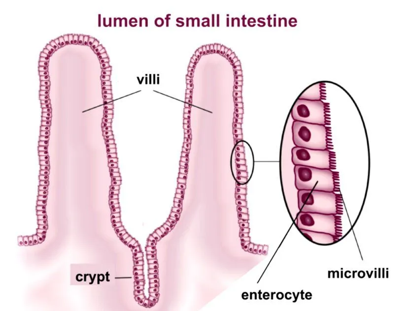
It should be noted that the small intestine has crypts that house stem cells, which serve to constantly replenish epithelial cells that are lost from the villi (intestinal villi are small, finger-like projections that extend into the lumen of the small intestine). It is of utmost importance that these stem cells are protected, as they contribute to the long-term maintenance of the intestinal epithelium.
Paneth cells are interspersed with these stem cells in the crypt base (typically 5 to 12 Paneth cells per crypt), not just sitting next to them. That close packing isn’t accidental: as the next section explains, the stem cells more or less depend on signals coming from their Paneth-cell neighbors to keep dividing.
Are Paneth Cells Found In The Large Intestine?
This is one of the most common follow-up questions about Paneth cells, and the textbook answer (“no, only in the small intestine”) is too clean. Here’s the more honest picture.
In a healthy adult human, Paneth cells are concentrated in the small intestine and reach their highest density in the terminal ileum. They are also present in small numbers in the proximal colon (the cecum and ascending colon), but they thin out as you move further down, and the rectum has essentially none. In mice and rats, the colon normally has no Paneth cells at all, which is part of why the “absent from the colon” shorthand stuck.
Why the difference? The colon has a much larger and more densely packed microbial population than the small intestine, but it manages that population with a different toolkit. The colonic epithelium leans heavily on a thick, double-layered mucus blanket secreted by goblet cells, plus a different family of antimicrobial peptides (mainly the β-defensins HBD1, HBD2, and HBD3, and the cathelicidin LL-37). The small intestine, with its thinner mucus layer and proximity to a much richer stream of nutrients (and the bacteria attracted to them), invests more heavily in Paneth-cell α-defensins to keep its crypt base aseptic.
There is one famous exception. In chronic intestinal inflammation, Paneth cells start showing up in places they normally wouldn’t, including the distal colon. This is called Paneth cell metaplasia, and it’s a histological marker of long-standing mucosal injury. It is seen in roughly 85% of pediatric ulcerative colitis cases and in most cases of Crohn’s colitis. It is the colon’s own (imperfect) attempt to reinforce a damaged barrier.
Paneth Cells And The Intestinal Stem Cell Niche
The intestinal lining is one of the most rapidly renewing tissues in the body. The cells that line your villi today will, for the most part, be sloughed off and replaced within about a week. That kind of churn requires a constant supply of fresh cells, and that supply comes from Lgr5+ stem cells parked at the very bottom of each crypt.
Here’s the catch. Stem cells, anywhere in the body, can’t maintain their stemness on their own. They need a “niche” of supporting cells that hand them the right chemical signals, like a backstage crew keeping a stage performer on cue. In a 2011 paper in Nature, Hans Clevers’ lab in Utrecht showed that, in the small intestine, that backstage crew is the Paneth cell. Paneth cells secrete Wnt3, EGF, TGF-α, and the Notch ligand Dll4, and the neighboring Lgr5+ stem cells sit and absorb that signaling soup.
So Paneth cells aren’t just hired security guards for the crypt. They’re also the local power supply for the stem cells. If you grow intestinal organoids (miniature crypts in a dish) and remove the Paneth cells, the stem cells struggle. Add them back, and the organoid grows happily again.
That said, the picture has been nuanced in more recent work. A 2025 review in Cellular and Molecular Gastroenterology and Hepatology describes Paneth cells as “dispensable yet irreplaceable” for the niche: if you genetically delete them, other epithelial cells can step up and provide some of the missing signals, but the crypt never works quite as well as it does with the original Paneth crew in place.
What Happens When Paneth Cells Go Wrong? (Crohnâs Disease)
One of the more striking discoveries of the last two decades is just how clearly Paneth-cell failure shows up in human disease, especially Crohnâs disease.
The strongest known genetic risk factor for Crohnâs is a mutation in a gene called NOD2, a bacterial sensor that lives inside Paneth cells (among others). Patients carrying the well-studied NOD2 1007fs variant produce noticeably less HD5 and HD6 in their ileal Paneth cells, which means the crypt base loses a chunk of its antimicrobial defense.
Two other Crohnâs-associated genes, ATG16L1 (an autophagy gene) and XBP1 (involved in handling ER stress), also disrupt Paneth-cell function. ATG16L1 T300A scrambles the way Paneth cells package and release their secretory granules. XBP1 mutations cause Paneth-cell stress and even cell death. The common thread is that all three converge on the same target: a Paneth cell that can no longer fire its full antimicrobial broadside when bacteria arrive. The result is a chronically inflamed ileum, the part of the gut where Crohnâs most often strikes.
This is a useful reminder that Paneth cells arenât just a textbook curiosity. They sit at the interface between a personâs genome and the trillions of microbes living a few microns away, and when that interface breaks, the consequences are real and clinical.
References (click to expand)
- Paneth Cells. Colorado State University, Pathophysiology of the Digestive System.
- Paneth Cells (histology image). University of Utah WebPath.
- Paneth Cells in intestinal homeostasis and inflammation. Yarovinsky Lab, University of Rochester Medical Center.
- Ouellette, A. J. (2016). Paneth cell α-defensins and enteric microbiota in health and disease. NIH/NCBI PMC.
- Sato, T. et al. (2011). Paneth cells constitute the niche for Lgr5 stem cells in intestinal crypts. Nature 469, 415–418.
- Schoofs, H. et al. (2025). Paneth Cells: Dispensable yet Irreplaceable for the Intestinal Stem Cell Niche. Cellular and Molecular Gastroenterology and Hepatology.
- Singh, R. et al. (2020). Metaplastic Paneth Cells in Extra-Intestinal Mucosal Niche Indicate a Link to Microbiome and Inflammation. Frontiers in Physiology.
- Wehkamp, J. et al. (2005). NOD2 (CARD15) mutations in Crohn’s disease are associated with diminished mucosal α-defensin expression. Gut.