Table of Contents (click to expand)
The corpus callosum is the brain’s largest white matter tract, a roughly 10 cm bundle of about 200 million axons that connects the left and right cerebral hemispheres. Its job is to shuttle motor, sensory and cognitive signals between the two sides, and damage to it can cause varying levels of impairment in mental and physical abilities.
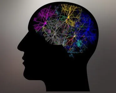
Our brain is the most complicated object we have encountered in the universe. It is powered by 86 billion neurons, yet uses just 20W of energy to get through the day. This 3-pound gooey organ sitting inside your skull is responsible for your upkeep; from the day you are born until the day you die. It stores your memories, develops your personality, and helps you read and understand what I’m currently writing; it is the container for every thought you have and is the processor of your interactions with the environment.
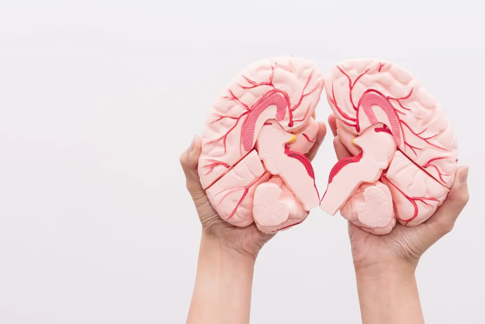
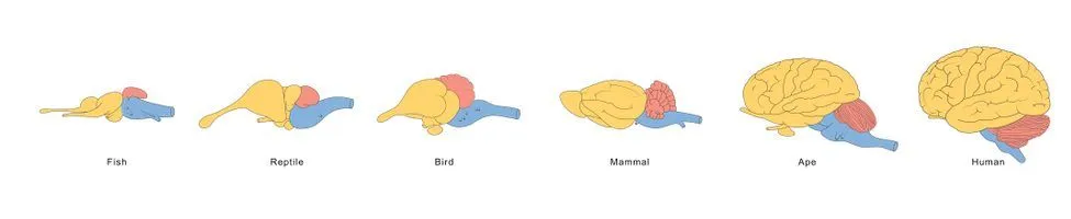
Understandably, this complex piece of machinery is divided into many different parts, which are responsible for all the varied things we do. The design of the brain is a testament to evolution, as higher functions and parts of the brain continually developed as animals evolved into higher levels of cognition.
Our brain is divided into two hemispheres. Each hemisphere has its own specific purposes and tasks, but many functions are dependent upon efficient communication between the hemispheres. The part of the brain that connects these two hemispheres and helps facilitate communication between the two is the corpus callosum. It is a very integral part of the brain, so damage to this region can cause severe impairments. Let’s take a look at the connector of the two sides of the brain and the consequences if it is underdeveloped or damaged.
The Connector Of The Hemispheres
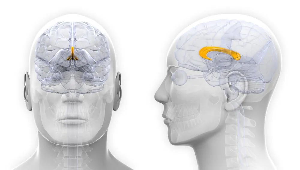
The corpus callosum sits beneath the cerebral cortex, deep in the brain. It is a thick, wide nerve tract made up of a flat bundle of commissural fibers, and it is one of the signature features of placental (eutherian) mammals, the group that carries its young in the womb until a relatively late stage of development; monotremes, marsupials and other vertebrates don’t have one. Roughly 10 centimeters (about 4 inches) long, it carries on the order of 200 million axons (estimates run as high as 300 million depending on the study), which makes it the largest white matter structure in the human brain. The basic shape of the corpus callosum is laid down between roughly the 8th and 20th weeks of gestation, growing from front to back, and it keeps thickening through the third trimester. Growth doesn’t stop at childhood either; MRI studies show the corpus callosum continues to enlarge and myelinate into the mid-20s.
Structure
The corpus callosum is the connector between the two cerebral hemispheres; it forms the floor of the longitudinal fissure, the deep groove that separates the two halves of the brain. Anatomists usually describe four named regions along its length, each a bundle of fibers linking different cortical areas: the rostrum, the genu, the trunk (or body), and the splenium. A narrow segment called the isthmus marks the transition between the trunk and the splenium.
The front end of the corpus callosum is called the genu (Latin for “knee”), which is tucked in towards the frontal lobes. The genu sits in front of the septum pellucidum, tapering in thickness as it curves backward and downward. The thin, beak-like extension where the genu tapers down is called the rostrum, which stretches toward the optic chiasm. The rear end of the corpus callosum is called the splenium, the thick, rounded portion that sits near the cerebellum, overlapping the tela choroidea of the third ventricle and the midbrain. It ends in a thick, convex border.
Connections And Functions
The fibers on either side of the corpus callosum radiate out through the white matter to reach the various parts of the cerebral cortex. A thin sheet of fibers from the splenium fans laterally and downward to form the roof and lateral wall of the lateral ventricle, extending into the temporal lobe; this layer is called the tapetum. Connecting the left and right temporal lobes is a job shared by the tapetum and the anterior commissure, a separate (and much smaller) interhemispheric bridge.
Myelin is the fatty substance that wraps nerve axons inside a neuron, insulating them and speeding up the rate at which information travels along the fiber. Myelination of the corpus callosum picks up speed in the first decade of life and continues at a slower pace through adolescence and into the third decade. The density of fibers in each sub-region, along with their size and degree of myelination, mirrors the functions of the brain regions they connect. Thick, heavily myelinated, fast-conducting fibers link the motor and visual areas. Thin, lightly myelinated fibers conduct more slowly and connect prefrontal and association areas.
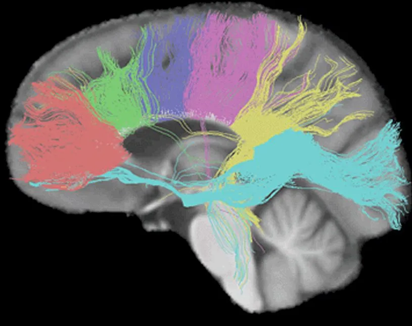
In a tractogram of the brain, the connections from the corpus callosum to various parts of the brain are seen in distinct colors. The fibers from the genu are seen in a reddish hue, fibers towards the premotor are in green, the sensory-motor fibers are purple, the parietals are pink, the temporals are yellow and those of the splenium are blue. The somatosensory (neurons that respond to changes on the exterior or interior of the body) information moving between the two halves of the parietal lobe and the visual cortex is communicated by the splenium.
Disorders And Medical Conditions
The corpus callosum is important for communicating information between the hemispheres. As seen above, it plays an important role in integrating and processing cognitive, motor and sensory information.
These functions can be affected when the corpus callosum is not properly formed, or in some cases, completely absent. Depending on the condition of the individual, the impact of this deformity could be subtle or severe. Research is being done to understand the various impacts of the disorder, as well as the differences and similarities between the following types:
- ACC or AgCC (Agenesis of corpus callosum): the complete or partial absence of the corpus callosum
- c-ACC (Complete agenesis of the corpus callosum): the complete absence of the corpus callosum
- p-ACC or Hypogenesis (Partial agenesis of corpus callosum): the partial absence of the corpus callosum
- Dysgenesis of the corpus callosum: The corpus callosum is completely present but malformed in some way
- Hypoplasia of corpus callosum: The corpus callosum is completely present, but is abnormally thin
The potential causes of these conditions range from injuries, toxic exposure, structural blockages (cysts), genetic factors or chromosome errors, metabolic disorders or other unknown factors. These abnormalities can be diagnosed via a CT scan or an MRI.
Some of the people diagnosed with one of the above conditions may be physically healthy and could have typical intelligence, but will show signs through subtle, yet significant symptoms. Those who are chronically suffering from a more serious condition could display additional brain abnormalities or medical and physical conditions that have a negative impact on their health and development to varying degrees.
A person suffering from such conditions could display the following characteristics:
- Epileptic seizures
- Cognitive impairments (learning disorders, abstract reasoning, problem-solving)
- Developmental delays in children (walking, talking, etc.)
- Sensory issues (visual, auditory, etc.)
- Behavioral disorders (ADD and ADHD)
- Sleep-related disorders (insomnia, sleep apnea, etc.)
- Clumsiness (poor motor coordination)
- Difficulty in understanding social cues
The disorders of the corpus callosum are a lifelong condition. Early intervention can help individuals, but continued therapy is required throughout their life. Research is being carried out to better understand the causes and impact of the impairment to the corpus callosum; this will enable families and professionals greater access to additional knowledge that would improve the lives of patients through more targeted support and intervention.
References (click to expand)
- Neuroscience For Kids - Hemispheres.
- Corpus Callosum - Developmental and Behavioral Pediatrics.
- Neuroanatomy, Corpus Callosum. StatPearls. NCBI Bookshelf.
- Corpus Callosum Agenesis. StatPearls. NCBI Bookshelf.
- Agenesis of the Corpus Callosum. BrainFacts.org (NINDS).
- Corpus callosum. Queensland Brain Institute, University of Queensland.