
Neither is simply better; it depends on the job. Electron microscopes magnify up to about 1,000,000x and resolve features below 1 nanometer, far beyond an optical microscope's roughly 200 nm limit and 1,000-1,500x magnification. But optical microscopes are cheaper, simpler, and can view living cells, which electron microscopes cannot.
Our eyes give us the vision we need to connect with the world around us. In the field of science, the discovery of the microscope has given many scientists the ability to look deep into the microscopic world and make some remarkable discoveries, such as that of the cell by Robert Hooke.
What Is A Microscope?
The term ‘microscope’ is derived from the Greek words for ‘small’ and ‘to view’. Thus, it is a device with which we can view small objects.
A microscope comes in two main types, depending on the construction: simple microscopes and compound microscopes.
A simple microscope uses only a single lens to obtain a magnified image. When using this microscope, the ultimate quality of the image is not very good, which is why it is not used for scientific studies.

On the other hand, a compound microscope is an arrangement that uses more than one lens to produce a magnified image of an object. It produces a high-resolution image of the object and is therefore ideal for scientific research purposes, laboratory and forensic purposes, and general industrial uses.
The two types of lenses used in the compound microscope are the objective lens and the eyepiece. The objective lens is placed facing the object to be examined, while the eyepiece is the lens through which the image is viewed. The eyepiece is also referred to as an ocular lens.
When the light passes through the objective lens from the object, a magnified, real image is produced. This image is further magnified by the eyepiece lens and a virtual image is produced when seen through the eyepiece, making greater detail visible.
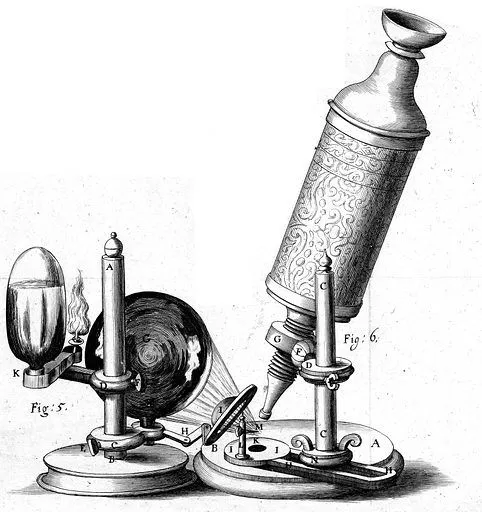
Microscopes are classified based on the kind of method they use to scan or examine the object.
What Is An Optical Microscope?
As the name suggests, an optical microscope uses light as the medium to scan objects, and through the system of lenses produces a magnified image. It is also called a ‘light microscope’. This is the most common type of microscope and finds use in various fields. They are relatively easy to construct and work with.
Since it generally uses visible light, the image produced can be directly perceived by the human eye.
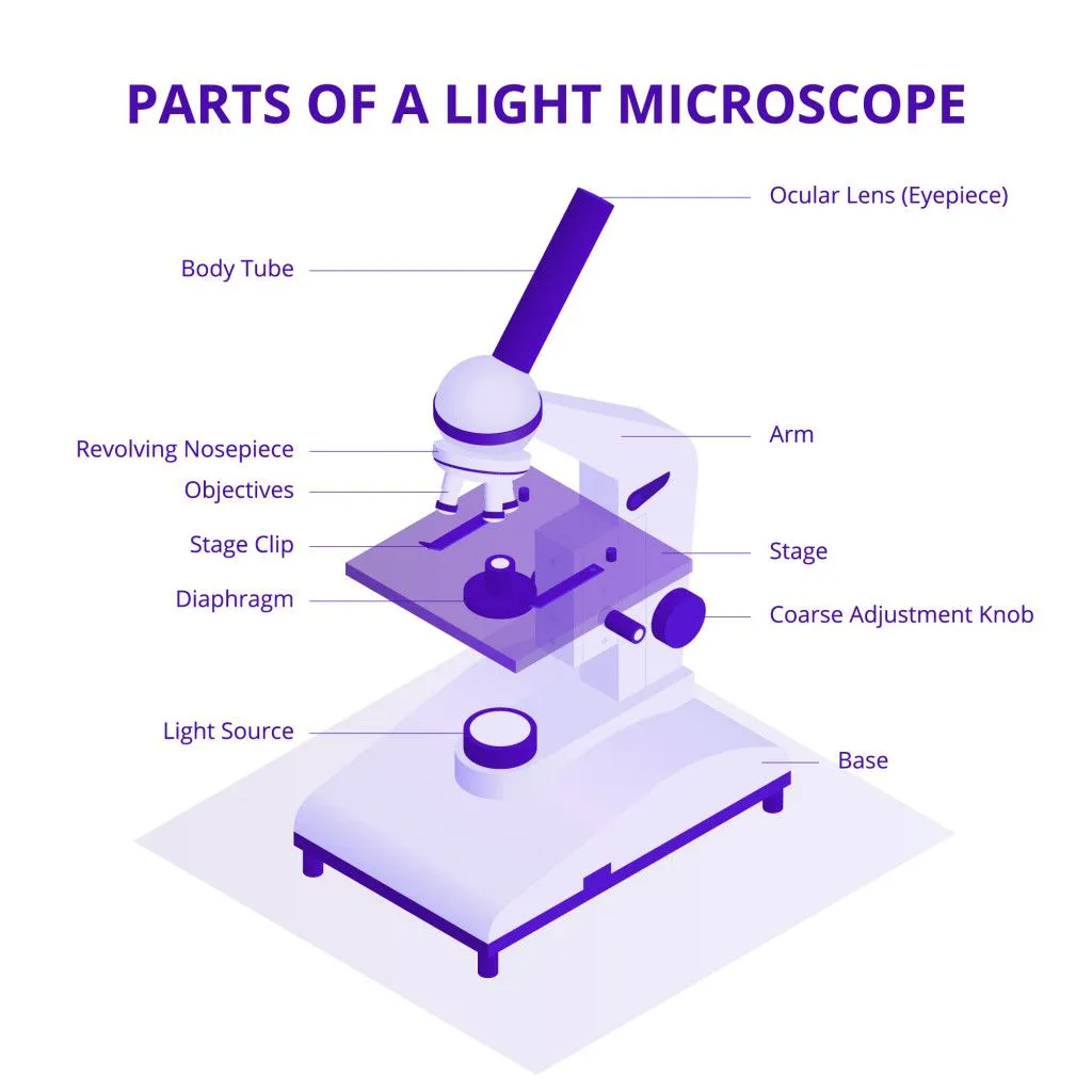
In a compound optical microscope, the brightness of the sample can be turned up or down, but this only changes how clearly we see the image, not how much fine detail it actually contains. The real limit on detail comes from the wavelength of visible light itself. Because light waves diffract, a light microscope cannot resolve two points that are closer together than about 200 nanometers (this is known as the Abbe diffraction limit, named after the German physicist Ernst Abbe, who described it in 1873). In practice, this caps the useful magnification of an optical microscope at roughly 1,000 to 1,500 times. Push the magnification higher and the image simply gets bigger and blurrier, without revealing anything new.
What Is An Electron Microscope?
An electron microscope uses an accelerated beam of electrons as the source of illumination of the specimen, and produces a very high-resolution image. Since the wavelength of a fast-moving electron is thousands of times smaller than that of visible light, the resolving power of the microscope is enormously high. The best modern electron microscopes can resolve features smaller than 0.1 nanometer (1 angstrom), fine enough to make out individual atoms, and can magnify a specimen up to a million times or more, compared to the roughly 200 nm and 1,000-1,500x of a light microscope.
Although the construction of this microscope is complicated, the image that it produces is incredibly detailed and not affected by atmospheric factors. Thus, this kind of microscope is used to study the internal structure and chemical composition of materials.
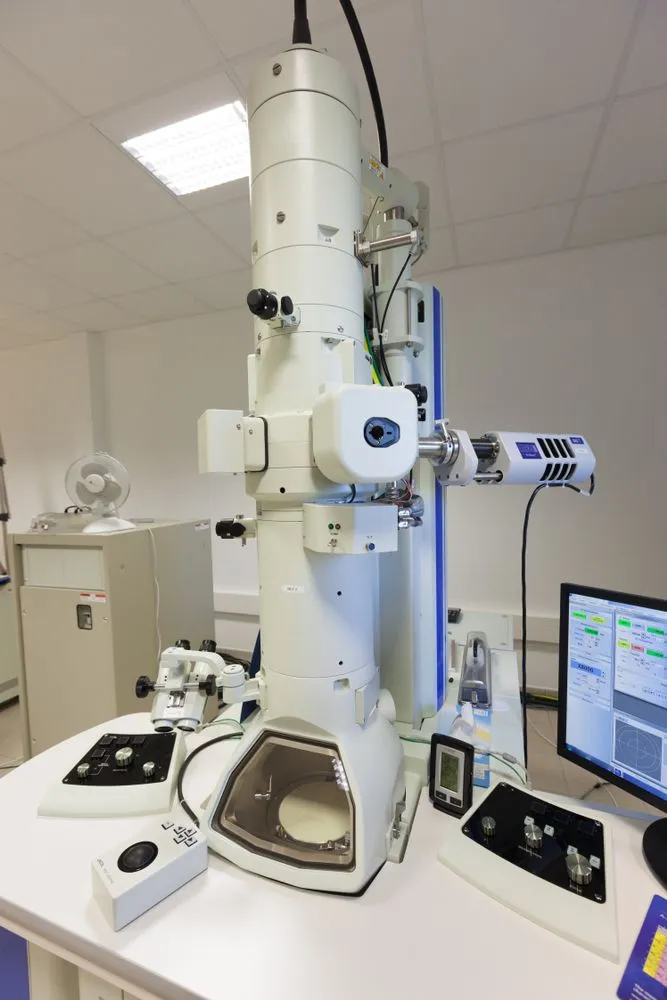
Electron microscopes are further divided into two main types: the Transmission Electron Microscope (TEM) and the Scanning Electron Microscope (SEM). In a TEM, the electron beam passes straight through an ultra-thin slice of the specimen, which reveals its internal structure in fine detail and gives the sharpest resolution of all. In an SEM, the beam instead sweeps back and forth across the surface, building up a striking three-dimensional picture of the specimen's exterior. Both are widely used in scientific and medical research.
Which Is Better: Optical Microscope Or Electron Microscope?
Both kinds of microscopes have their fair share of advantages and disadvantages. It is completely dependent on the usage and tasks to which they are assigned.
While the electron microscope has higher resolving power and produces higher resolution images than the optical microscope, it requires a vacuum to work. Because the sample sits in that vacuum and is usually fixed, dehydrated, and coated with a thin metal layer before imaging, an electron microscope cannot view living cells, only dead and carefully prepared ones. The specimen preparation and the image production also take far longer than in an optical microscope, and the instruments themselves are large and expensive, often costing many times more than a quality light microscope.

On the other hand, the optical microscope is cheaper, lighter, and far easier to use, needs little or no sample preparation, and (crucially) can image living, moving cells in their natural watery state, often in full color. Its main drawback is that the image simply cannot capture the fine internal detail that an electron microscope can, because of the diffraction limit set by the wavelength of visible light.
One must choose wisely between the two, keeping in mind the tasks you need to complete.
Future Developments In Microscopy
The neat divide between the two families is starting to blur. For a long time, the 200 nm diffraction limit was treated as an unbreakable wall for light microscopes. Then came super-resolution fluorescence microscopy, a clever set of techniques (such as STED and PALM) that sidestep the Abbe limit and let a light microscope resolve detail down to a few tens of nanometers, all while keeping the sample alive. The breakthrough was considered so important that Eric Betzig, Stefan Hell, and William Moerner shared the 2014 Nobel Prize in Chemistry for developing it.
At the same time, microscopes of every kind are increasingly paired with sophisticated software and machine-learning algorithms that sharpen noisy images, reconstruct three-dimensional models, and pick out features the human eye would miss. Resolution, speed, accuracy, and sensitivity remain the key areas of focus in microscopy, both today and in the near future.
Conclusion
Each little step, each thought towards a broader vision, improves the possibilities of evolution. When we have the curiosity to look beyond what our physical senses have to offer, revolutionary discoveries inevitably occur!
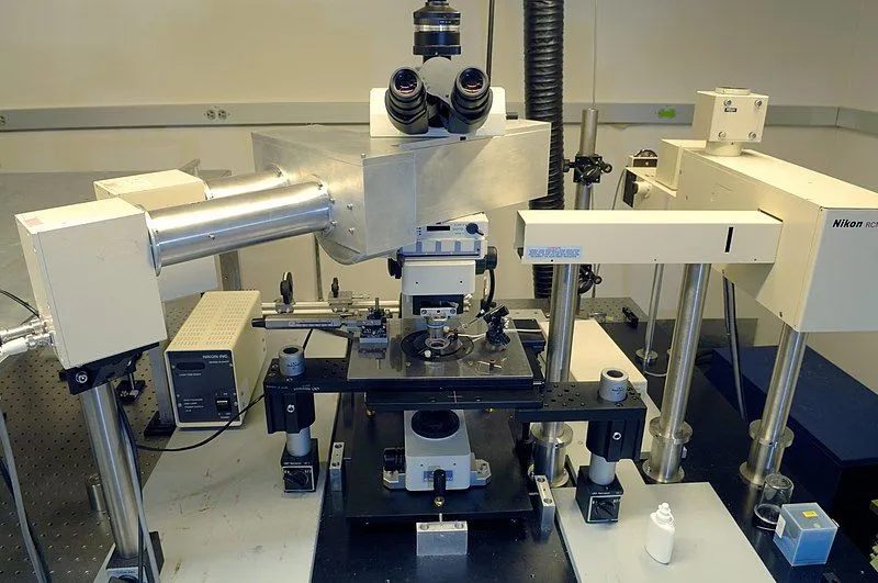
The microscope is one such discovery that gave the correct tool to scientists and researchers, who then opened the closed doors to the microscopic world. With further advancements in Science and Engineering, the unknown areas of the microscopic world will be even more accessible to us.
References (click to expand)
- What Is an Electron Microscope (EM) and How Does It Work?. The United States Department of Veterans Affairs
- Microscope. Encyclopaedia Britannica
- The Evolution of the Microscope - JSTOR Daily. JSTOR
- MW Davidson. OPTICAL MICROSCOPY - Zeiss Campus. Florida State University
- Types of electron microscope - Science Learning Hub. sciencelearn.org.nz
- Electron microscope. Encyclopaedia Britannica
- The Nobel Prize in Chemistry 2014 (super-resolved fluorescence microscopy). NobelPrize.org