
The basilar membrane is a structural element inside the cochlea that separates two of its three fluid-filled chambers (the scala media and the scala tympani). It transforms sound waves into electrical impulses transmitted to the brain, and helps identify the frequency profile of incoming sound by acting like a bank of band-pass filters.
The basilar membrane is an important component of the inner ear and is located inside the cochlea, which is moved by sound waves that fall on the ear. This delicate structure is critical for our sense of hearing.
The Inner Ear
Some of you may already know that the ear can be divided into three parts: the outer ear, middle ear and inner ear, all of which work in perfect synchronization to help us hear what is happening in the outside world.
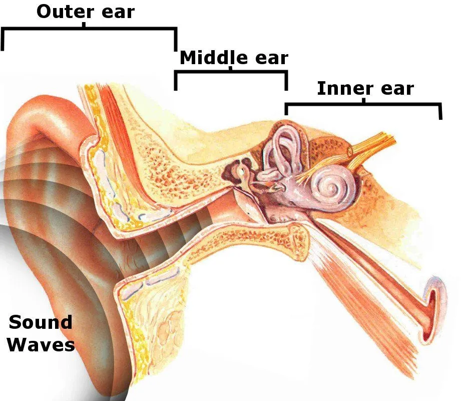
While the first two, i.e., the outer ear and middle ear, are filled with air, the inner ear is filled with fluid. As its name signifies, the inner ear is the innermost segment of the ear, and consists of a maze of passages and tubes (called the labyrinth). It consists of the cochlea, the balance mechanism, the vestibular nerve and the auditory nerve.
The sounds that we hear fall on our ears like waves, which then pass through the outer and middle ear to finally reach the cochlea. The cochlea is a very important part of the inner ear; it looks like a wound-up hose or a snail shell and contains sensory cells that can detect sound.
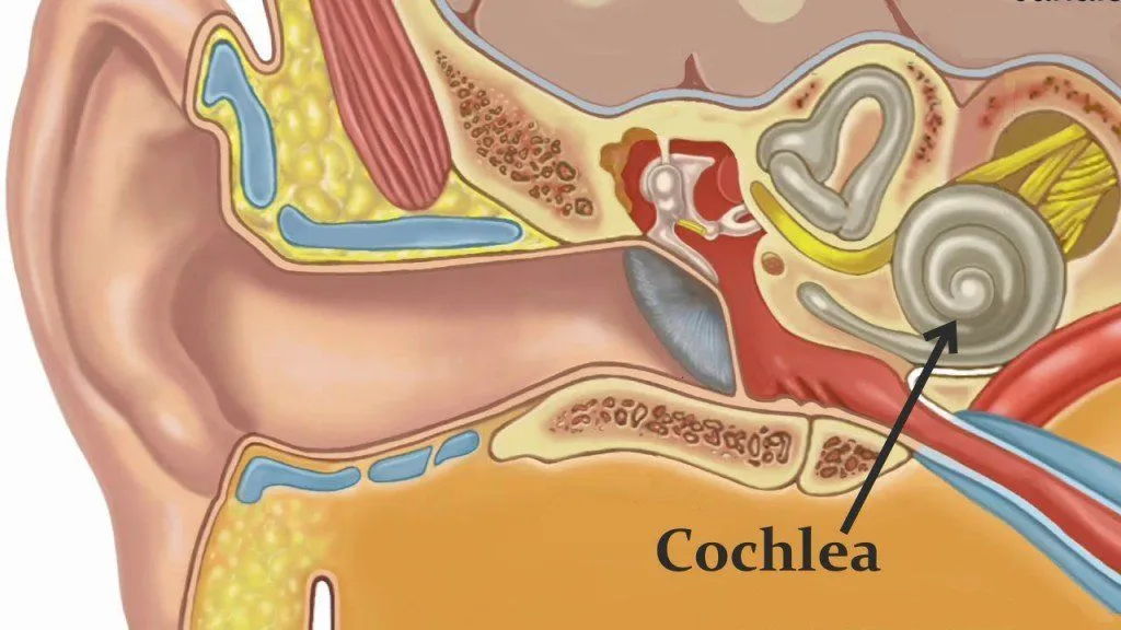
When sound waves reach the cochlea, they are transformed into electrical impulses. These are subsequently transmitted to the brain, which decodes these impulses and ultimately registers it as a sound.
What Is The Basilar Membrane?
The cochlea is essentially a long, coiled tube divided into three parallel, fluid-filled chambers: the scala vestibuli (top), the scala media (middle), and the scala tympani (bottom). Two membranes do the dividing. Reissner's membrane separates the scala vestibuli from the scala media, and the basilar membrane separates the scala media from the scala tympani.
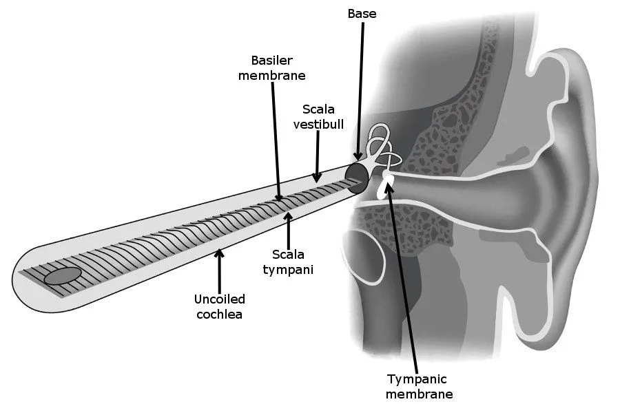
The basilar membrane does not have a uniform thickness or stiffness, because how thick or stiff it is at a given point on its length determines its characteristic frequency (CF), which is a particular frequency value at which the basilar membrane is most sensitive to sound vibrations.
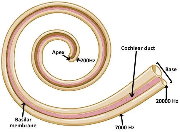
The basilar membrane is the narrowest (0.08-0.16 mm) and most stiff at the base of the cochlea, and the widest (0.42-0.65 mm) and least stiff at the apex. Note that low-frequency sounds localize near the apex of the cochlea, while high-frequency sounds localize near the base. This sort of non-uniformity in the structure of the basilar membrane facilitates high-frequency sounds moving only a small region of the basilar membrane near the stapes (a small bone in the inner ear), whereas low frequencies cause almost the entire membrane to move.
The basilar membrane has roughly 15,500 sensory hair cells attached to its surface: about 3,500 inner hair cells in a single row, and around 12,000 outer hair cells arranged in three rows. These are not the hair that we all have on our heads, but rather a type of mechanoreceptor, meaning that they are activated by movement. These hair cells are exclusively moved by incoming sound waves. The basilar membrane, the tectorial membrane and the hair cells are jointly called the Organ of Corti.
Functions Of The Basilar Membrane
The movement of the basilar membrane is essentially what allows humans to hear through their ears. It works like this: the movement of the basilar membrane causes the hair cells’ cilia to brush gently against the surface of the tectorial membrane. This sort of bending movement prompts the hair cells to fire a neural impulse, telling the brain that a sound wave was detected.
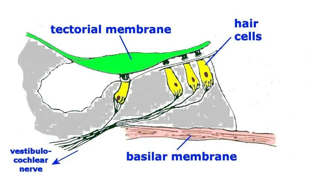
The brain then decodes that neural signal and voila! We hear things!
Apart from serving this critical function of helping us hear, the displacement of the basilar membrane helps to identify the profile of the sound impinging on the ear by acting like a series of band-pass filters. Using a technique called laser interference, these filtering characteristics of the basilar membrane can be studied in detail.
Basilar membrane is just one of many tiny components present inside the ear which work together in perfect harmony to make us hear what the world has to say to us!
Base vs Apex Of The Cochlea: How The Basilar Membrane Sorts Frequencies
Have you ever wondered how a single membrane can tell the rumble of a bass drum apart from the shrill of a whistle? The trick lies in the fact that the basilar membrane is not the same all the way along its length. At the base of the cochlea, near where the sound first enters, it is narrow and stiff. By the time you reach the apex (the very tip of the coiled snail shell), it has become wide and floppy. That single change in mechanics is what lets us hear across roughly 10 octaves, from about 20 Hz up to 20,000 Hz.

Here is the part that trips a lot of people up, so it is worth stating plainly. The stiff base responds best to high-frequency sounds, while the flexible apex responds best to low-frequency sounds. Think of it like the strings of a guitar: a short, tight string sounds a high note, while a long, slack one sounds a low note. Because the stiffness of the membrane increases steadily from the apex toward the base, every spot along its length has its own preferred pitch, called its characteristic frequency.
This orderly arrangement of pitch along the membrane is what scientists call tonotopy (or tonotopic organization). When a sound enters the ear, it sets off a traveling wave that ripples from the base toward the apex, growing in size until it peaks at the one location tuned to that exact frequency, and then dies away. The Hungarian biophysicist Georg von Bekesy won a Nobel Prize for working this out by watching these waves move along the cochlea. In psychology classes this idea is taught as the place theory of pitch perception: the brain figures out the pitch of a sound largely from where along the basilar membrane the vibration is strongest. So a query like "base vs apex of cochlea" really comes down to one neat rule: base equals high notes, apex equals low notes.
Tectorial Membrane vs Basilar Membrane: What's The Difference?
If you have read this far, you have met two different membranes, and it is easy to get them muddled. They sit close together and work as a team, but they do very different jobs. The basilar membrane is the floor that the hair cells stand on; it is the structure that separates the scala media from the scala tympani and physically vibrates in response to sound. The tectorial membrane is a separate, gel-like ribbon of tissue that hangs over the top of the hair cells like an awning.

The magic happens in the gap between the two. When the basilar membrane bobs up and down, the hair cells riding on it move too, while the tectorial membrane above shifts in a slightly different way. This mismatch produces a sideways shearing motion that bends the tiny hair bundles (stereocilia) on top of the hair cells. That bending pries open channels at the tips of the stereocilia, the hair cell fires, and a nerve signal is sent off to the brain. No shearing, no hearing.
The two membranes also treat the two kinds of hair cells differently. The tallest stereocilia of the outer hair cells are actually embedded right into the underside of the tectorial membrane, so they are dragged directly as it moves. The inner hair cells are choosier: their stereocilia stand free in the fluid and are not anchored to the tectorial membrane, so they are nudged by the movement of the surrounding fluid instead. Together with the hair cells, the basilar and tectorial membranes make up the Organ of Corti, the true sensory engine of hearing. In short, the basilar membrane does the vibrating, the tectorial membrane provides the surface to shear against, and the hair cells turn that tiny tug into the sound you experience.
References (click to expand)
- Cochlea and Vestibular System - courses.washington.edu:80
- The Organ of Corti in the Inner Ear.
- Science, Maths & Technology - OpenLearn.
- The Basilar Membrane and Fourier Analysis.
- Hearing and Hair Cells - www.neurophys.wisc.edu:80
- Physiology, Cochlear Function. StatPearls. NCBI Bookshelf.
- Anatomy, Head and Neck, Ear Organ of Corti. StatPearls. NCBI Bookshelf.
- The Inner Ear. Neuroscience, 2nd edition. NCBI Bookshelf.
- How Do We Hear? National Institute on Deafness and Other Communication Disorders (NIDCD).
- Georg von Bekesy. Encyclopaedia Britannica.