Living cells hum. Their membranes vibrate a few nanometers up and down about a thousand times per second, and yeast cells, for example, give off a tone near 880 Hz, roughly the pitch of a dial tone. The vibrations are driven by motor proteins like kinesin and myosin shuttling cargo around inside the cell. The study of these cellular sounds is called sonocytology, a field launched by UCLA’s Jim Gimzewski and Andrew Pelling in 2004 using an atomic force microscope.
The inner voice that often tells you to take the leap, quit the job, confess to your crush and give voice to your random violent thoughts may actually be your cells singing a symphony.
Well, not really, but your cells do sing and they might be able to give opera singers a run for their money.
Researchers in the year 2004 discovered that cells vibrate; when those vibrations are amplified, they sound like squeals. Also, each individual cell is thought to vibrate uniquely. Scientists argue that decoding these squeals could help us recognize the state of a cell and thus predict the arrival of a disease. This study of the sounds of a cell is called sonocytology (“sonos” ~ sound, “cytology” ~ study of cell structure and function).
Discovery Of Sonocytology
Jim Gimzewski, a UCLA professor and physicist, along with graduate student Andrew Pelling, were the researchers to discover cellular sounds. Jim’s interest was piqued in the matter when Carlo Ventura, a medical researcher, informed him of his ongoing study on stem cells and their implantation in heart attack patients. The stem cells would need to be implanted at precisely the right moment (just when they start beating) in order to help heart patients. Jim asked Carlo to send him a few cells, stating that he would listen to them in order to find the exact moment in their development when they start beating.
He was also curious to see if all other cells beat, or if this was unique to the cells of the heart. Furthermore, if all cells indeed throbbed like heart cells, would the vibrations be sufficient to produce a detectable sound?
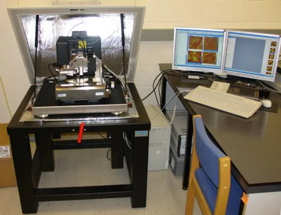
Being an expert in the field of instrumentation, Jim knew that the sound a cell produced wouldn’t be audible to the naked ear, but could be detected using a highly sensitive apparatus. The pair therefore made use of an atomic force microscope (AFM) to conduct their studies and hear the cells.
Due to the tragic circumstances of 9/11, the stem cells never arrived. However, Jim couldn’t wait to hear the song of the cells and instead decided to experiment on yeast cells that he obtained from his colleagues at UCLA. Yeast cells are often at the heart of many biological studies due to their rapid reproduction rates and the presence of fewer genes.
Gimzewski and Pelling’s studies in a small soundproof room in the basement of UCLA’s Young hall eventually led them to establish the field of sonocytology in the year 2004. That being said, the field of sonocytology is still vastly unexplored and even lacks a Wikipedia page of its own!
Atomic Force Microscope (AFM)
Before we go into further detail about the sounds of a cell, let’s take a brief look at the apparatus these researchers used to detect these sounds.
An atomic force microscope is a highly sensitive scanning probe capable of creating images of extremely tiny objects. This includes objects smaller than a nanometer (1 nanometer = 10–9 meter). The human hair itself is about 80,000 – 100,000 nm wide, and an AFM is capable of imaging objects much smaller than that.
The AFM consists of a cantilever beam with an extremely tiny tip (typically made of silicon or silicon nitride) affixed at one end. Much like a record player, the tip touches the surface to be imaged, while an xyz drive moves the cantilever beam across the surface. The tip is deflected every time it detects a ‘hill’ or ‘valley’ on the surface. This deflection is picked up by a laser being reflected from the cantilever beam. For every deflection of the tip, the laser is also forced to change its direction. The final result is a detailed image with every hill and valley mapped out on the surface of the nanoscopic object.
What Makes Cells Sing?
Coming back to our singing cells, Gimzewski & Pelling’s first experiment on the yeast cells shocked the world. The AFM recorded regular oscillations arising from the cell wall. The cell wall raised and fell by three nanometers (amplitude of the sound wave) and repeated this nearly 1,000 times per second (frequency of the sound wave). Using conversion software, the pair managed to turn the AFM image into a sound that could be played back through speakers. The result was a tone of about 880 Hz.
However, Gimzewski & Pelling were worried that the sound may have been due to some inner machinery of the microscope. The pair therefore ran even more tests on the yeast cells to confirm their initial findings.
They soaked the cells in rubbing alcohol (known to break the cell membrane and lead to cell death) to check if there was any change in the pitch of the sound. To quote Jim himself, rubbing alcohol made the cells scream at first, before silencing them to death. After replicating their experiments in various conditions, such as different chemical media and temperatures, and on different types of cells, they concluded that each cell produces its own unique sound. Their experiments also suggested that the sounds may be arising from inside the cells.
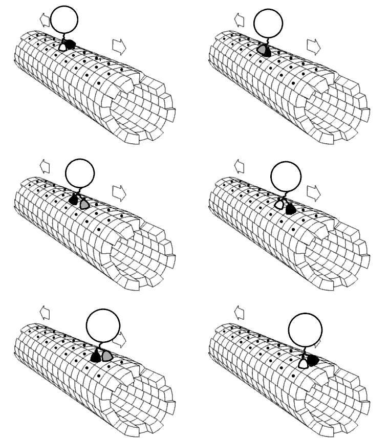
Jim believes the sound originates from the movement of molecular motors. Molecular motors are biological machines responsible for the transportation of materials inside a cell. They are essentially protein molecules (and are also called motor proteins) that utilize chemical energy from ATP hydrolysis to move along the cytoplasm of cells. Some of these motor proteins are responsible for carrying cell organelles like mitochondria, Golgi stacks, etc. to their individual locations in the cell, while others set the stage for phenomena like cell division, muscle contraction, etc. Examples of motor proteins are Kinesin, Myosins, Dynein, etc.
Motor proteins are connected to the cytoskeleton (responsible for maintaining the structural integrity and shape of a cell), which in turn is connected to the cell membrane. The movement of motor proteins is therefore believed to be carried through to the cell membrane, causing it to oscillate, through the cytoskeleton.
According to Jim’s calculation, the energy required for the motor proteins to move and produce sound is the same. In support of Jim’s theory, motor molecules take roughly 1000 steps per second, which was also the frequency of the sound the AFM recorded. The unified movement of the motor proteins inside a cell is therefore believed to be the reason behind the sounds of a cell!
What Do Singing Cells Actually Sound Like?
So what does a cell's song actually sound like to human ears? On its own, nothing at all. A cell's surface only rises and falls by around three nanometers, and a vibration that tiny moves far too little air for us to hear it directly. Only after the AFM's readings are amplified and converted into audio do they land inside our hearing range. A healthy yeast cell then comes through as a steady tone of roughly 880 Hz, close to the pitch of an old telephone dial tone.

That note is not fixed, though. Gimzewski and Pelling found that a cell's frequency shifts with its temperature and metabolism, so the same cell does not always sing at exactly the same pitch. The sharpest change came at the moment of death. When the pair sprinkled rubbing alcohol on a yeast cell, its pitch shot upward into what they described as a scream, and once the cell had died the singing stopped. What remained was only a low, rumbling sound that Gimzewski suspects comes from random atomic motion rather than from anything the cell is actively doing.
The recordings were striking enough to cross over into art. In 2004, media artist Anne Niemetz teamed up with Andrew Pelling to create The Dark Side of the Cell, an immersive sound installation that premiered at the Los Angeles County Museum of Art. Visitors stood in a darkened room and listened to the amplified sounds of living and dying cells played through speakers, in what is described as the first musical composition built entirely from the voices of real cells.
Do Human Cells Sing Too?
Yeast made a convenient first subject, but the obvious question is whether our own cells hum as well. Gimzewski and Pelling went on to test several kinds of mammalian cells, the same broad family that our bodies are built from, and reported that these sing too. Bone cells, for example, were recorded at a lower pitch than yeast, though the researchers admitted they could not yet explain why different cell types settle on different notes.

If mammalian cells really do have their own voices, then in principle the tissues of the human body are quietly humming as well, each cell type with its own signature note. It is worth keeping expectations grounded, though. Much of this early sonocytology work was never published in a peer-reviewed journal, and the field remains small and largely unexplored. For now, the idea that your own cells are singing is a genuinely tantalizing possibility at the frontier of biology rather than a settled scientific fact.
Final Words
With every new piece of research come new critics. Some believe that there aren’t enough molecular motors to generate an audible sound, so ribosomes could be the ones making the cells sing. However, scientists are now more focused on how cellular sounds could be used for the public benefit, rather than finding their definitive origin.
The most interesting application of cellular sounds and the field of sonocytology proposed so far is the detection of diseases, such as cancer. As mentioned earlier, different types of cells make different types of sounds. This also includes mutant and cancerous cells. Cancerous cells are known to have a softer cell membrane, so if everything goes to plan, using the sounds of a cell and the AFM, the checking of a single cell could be enough to diagnose cancer. Also, sonocytology could help to create a library of sounds generated by cells affected with various diseases to help predict their arrival or development!
References (click to expand)
- Science Notes 2005: Getting the Vibe. The University of California, Santa Cruz
- Fundamental Theory of Atomic Force Microscopy. The Georgia Institute of Technology
- Sounds of Cells Dividing - Gerri Ondrizek. Reed College
- (2002) Molecular Motors - Molecular Biology of the Cell - NCBI. The National Center for Biotechnology Information
- Signal Discovery? Smithsonian Magazine
- The Dark Side of the Cell - Interalia Magazine