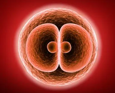
There are two main types of cell division: mitosis and meiosis. Mitosis produces two identical diploid daughter cells (46 chromosomes in humans) for growth and repair. Meiosis involves two divisions and produces four genetically unique haploid gametes (23 chromosomes) for sexual reproduction (sperm and eggs).
We are born as little babies and grow up into an adult, but what is the process behind this growth? In addition, we all know about sperms and eggs, so do they have the same process to produce them or is it different? Well, cell cycle has two ways about it. First is Mitosis and the second is Meiosis. Mitosis is what gets us from a zygote to a full-grown adult while meiosis makes gametes or sex cell, i.e. sperm and egg.
Cell Cycle: Interphase And Mitotic Phase
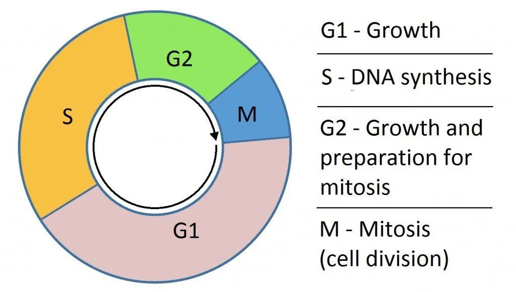
Before we start with understanding these two processes, we need to know a few things. First term is the cell cycle. The cell cycle is a sequence of events that happens before the cell finally divides and forms two daughter cells. It has two main phases known as Interphase and Mitotic phase. The interphase is divided into three phases, which are G1 (First Growth), S (DNA Replication) and G2 (Second Growth). During Interphase, the cell is preparing for the mitosis.
Haploid Vs Diploid
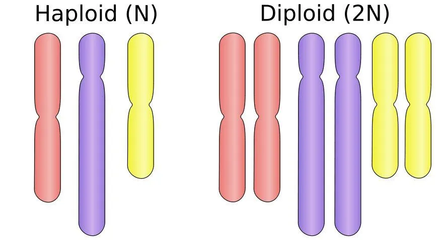
Next are haploid and diploid. Haploid refers to a single copy of chromosomes in a cell while diploid is for two copies of the chromosomes. Human cells are diploid. We get one copy from our father and one from our mother. However, the gametes or sex cells are haploid.
Finally, we need to know the cell organelle which plays an important part in mitosis/meiosis. They are centrosomes. Centrosomes are made up of microtubules and centrioles. Microtubules form the spindle fibers and centrioles help organize the spindle into proper formation.
Now we can pay to attention to the cell division process. Both of them have the same phases, namely, prophase, prometaphase, metaphase, anaphase, telophase and cytokinesis.
What Is Mitosis – The Equational Division
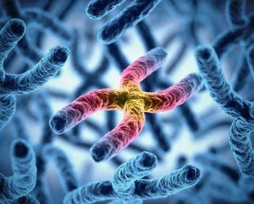
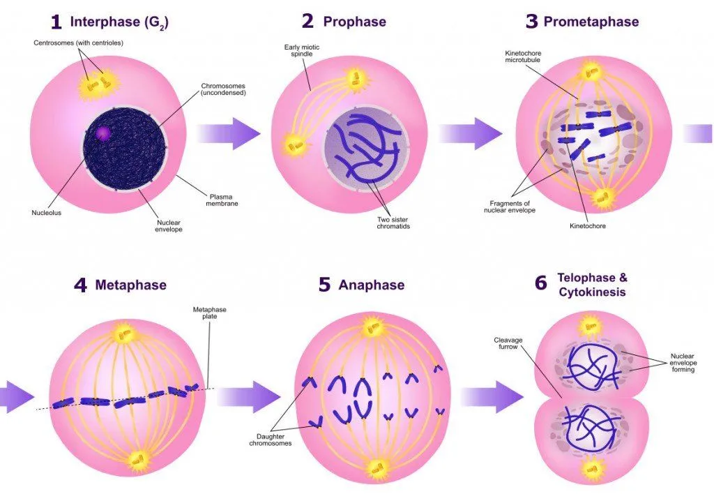
The cell grows and prepares itself for the mitotic division in its interphase. This involves duplication of centrosomes, chromosomes and other cell organelles. The chromosomes duplicate and form sister chromatids which are joined at the center via centromeres. These chromatids will split during mitosis and two daughter cells will get one each. This replication and equal division of chromosomes is the reason for the term ‘equational division’.
- Prophase – The chromatin begins to condense and the centrioles move towards the opposite ends. The microtubule fibers cross the cell to form mitotic spindle.
- Prometaphase – It begins with the dissolution of nuclear membrane. Proteins attach to the centromeres and form Kinetochores and the fibers attach to them. The kinetochores are one per chromatid. They are at the center of chromosomes. Once attached, the chromosomes start to move towards the center.
- Metaphase – The spindle fibers help in aligning the chromosomes in a straight line at the centre of the cell. This line is called the metaphase plate. Such an alignment helps in proper separation of chromosomes and ensures that each daughter cell gets one copy of the chromosomes.
- Anaphase – In this phase, the chromatids separate at the kinetochore. After separation they begin moving to opposite ends. The movement is a result of kinetochore action along the fibers and the physical interaction of the polar microtubules.
- Telophase – The chromosomes reach the opposite ends and expand. Formation of nuclear membrane also begins. Finally the spindle fibers disappear and cytokinesis begins.
- Cytokinesis – This procedure is the separation of the two daughter cells. A fiber ring made up of actin (protein) forms around the centre of the cell and pinches the cell to make it split into two diploid (46 chromosomes) daughter cells.
When mitosis goes wrong, the consequences can be severe. Errors in chromosome separation can lead to cells with abnormal chromosome numbers (a condition called aneuploidy), which is a hallmark of cancer cells. Our cells have built-in quality control mechanisms, including the spindle assembly checkpoint (SAC), which ensures chromosomes are properly attached before separation proceeds. A 2024 study discovered a "Mitotic Stopwatch" complex that monitors how long mitosis takes; if the process stalls (normal mitosis takes about 30 minutes), this molecular alarm prevents the potentially dangerous cell from continuing to divide.
Which Is The Shortest Phase Of Mitosis?
If you line up the stages of mitosis by how long they last, one of them barely registers. Anaphase is the shortest phase of mitosis. A typical human cell spends roughly 30 minutes in mitosis overall, but anaphase itself is finished in just a few minutes.

The reason comes down to how little anaphase actually has to do. By the time the cell reaches it, all the demanding preparation is already complete: the chromosomes are condensed, the nuclear membrane has broken down, and every chromosome is lined up on the metaphase plate with spindle fibers hooked onto its kinetochores. Anaphase is simply the moment the sister chromatids split at their centromeres and get hauled to opposite poles. In most animal cells the chromatids travel toward the poles at roughly one micrometre per second, so the separation is over almost as soon as it begins.
Prophase and prometaphase, by contrast, involve the slow, careful work of condensing the chromosomes, dismantling the nuclear envelope and assembling the spindle, which is why they eat up much more of mitosis. Anaphase inherits a cell that is already set up and ready, so it flashes past. The same logic carries over into meiosis, where the matching separation steps are similarly brief.
What Is Meiosis – The Reductional Division
Meiosis is used to produce gametes or sex cells. Even though the phases involved are similar to that of mitosis, there are a few things that are unique. The process takes place in two parts.
- Meiosis I – This process gives us two haploid daughter cells.
- Prophase I – After DNA replication and formation of sister chromatids, homologous chromosomes pair up and form synapsis. These paired chromosomes are known as bivalents. Chiasmata is formed which causes genetic recombination as the genetic content is exchanged. This step is important as it leads to genetic variability.
- Prometaphase I – The nuclear membrane disappears and kinetochore is formed. Each chromosome has one kinetochore instead of each chromatid. The spindle fibers attach and the chromosomes move to the middle.
- Metaphase I – The bivalents align in a straight line at the metaphase plate. The orientation of chromosomes is random as any of the parental homologue can be can be on either sides.
- Anaphase I – The chromosomes, each with two chromatids separate and move to the opposite poles.
- Telophase I – The nuclear envelopes appear and cytokinesis is initiated. This gives us two haploid daughter cells but with two sister chromatids.
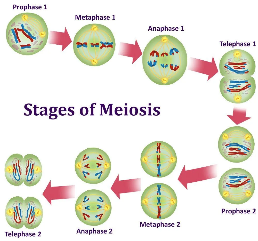
In meiosis two, there is no S phase i.e. no duplication of chromosomes. The process is similar to mitosis as the two sister chromatids are separated and the two haploid daughter cells form four haploid (23 chromosomes) cells with a single chromatid. This reduction in chromosome number is the reason for naming meiosis as ‘reductional’ division.
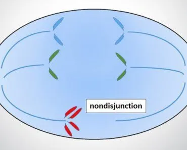
When chromosomes fail to separate properly during meiosis (a phenomenon called nondisjunction), the resulting gametes have an incorrect number of chromosomes. If such a gamete participates in fertilization, the resulting embryo will have too many or too few chromosomes. The most well-known example is Down syndrome, caused by an extra copy of chromosome 21 (trisomy 21), resulting from nondisjunction during meiosis.
The Phases Of Meiosis II (And How It Differs From Meiosis I)
Meiosis I leaves you with two haploid cells, but each chromosome inside them still carries two sister chromatids. Meiosis II is the second division that finally pulls those chromatids apart. The important thing to notice is that there is no S phase beforehand, so the DNA is not copied again. The short pause between the two divisions is called interkinesis, and because it skips replication, the cell moves straight into a division that closely resembles mitosis. Here is what happens, step by step:

- Prophase II – The chromosomes condense once more, the nuclear envelope breaks down again, and a fresh spindle is assembled in each of the two cells.
- Metaphase II – The chromosomes line up single file along the metaphase plate, with the sister chromatids attached to spindle fibers reaching in from opposite poles.
- Anaphase II – The centromeres split and the sister chromatids are finally separated, moving to opposite ends of the cell as individual chromosomes.
- Telophase II and cytokinesis – Nuclear envelopes reform, the chromosomes decondense, and each cell pinches in two during cytokinesis. The end product is four genetically unique haploid cells.
The key difference between the two divisions is what gets separated. In meiosis I, the homologous chromosomes (the matching pair, one inherited from each parent) are pulled apart, which is what halves the chromosome number and earns meiosis I the name reductional division. In meiosis II, it is the sister chromatids of each chromosome that separate, exactly as they do in mitosis. So anaphase I moves whole chromosomes to opposite poles, while anaphase II moves single chromatids. In effect, meiosis II is a tidy, mitosis-style division that finishes the job meiosis I began.
Key Differences: Mitosis vs Meiosis
- Number of divisions: Mitosis involves one division; meiosis involves two.
- Daughter cells: Mitosis produces 2 identical diploid cells (46 chromosomes); meiosis produces 4 genetically unique haploid cells (23 chromosomes).
- Genetic variation: Mitosis produces clones; meiosis introduces variation through crossing over and independent assortment.
- Purpose: Mitosis is for growth and repair; meiosis is for producing sex cells (gametes).
- Where it occurs: Mitosis occurs in somatic (body) cells; meiosis occurs only in reproductive organs (ovaries and testes).
References (click to expand)
- Phases of Mitosis - Khan Academy.
- Phases of Meiosis - Khan Academy.
- or or - oli.cmu.edu
- http://web.archive.org/web/20200805131323/http://www.tusculum.edu/faculty/home/ivanlare/html/genetics/meiosis-master.html
- Cell division quality control 'stopwatch' uncovered. ScienceDaily (2024).
- Memories of mitosis: Detecting defects during cell division. ScienceDaily (2024).
- The Process of Meiosis: Meiosis II - Biology LibreTexts.
- Anaphase - Molecular Expressions, Florida State University.