Table of Contents (click to expand)
- mRNA: The Starting Point Of Translation
- tRNA: The Assembly Point Of Translation
- Wobble Hypothesis: Why 20 Amino Acids Have 64 Codons?
- How Many Codons Code For One Amino Acid?
- How Do You Read The Genetic Code To Find An Amino Acid Sequence?
- The Significance Of The Wobble Hypothesis
- What's The Advantage Of Having Multiple Codons?
The genetic code is "degenerate": its 64 codons (each a triplet of mRNA bases) code for just 20 amino acids, so most amino acids are specified by more than one codon. This works because of wobble base pairing. At the third position of the codon, the pairing with the first base of the tRNA anticodon is loose, so a single tRNA can recognize two, three, or even four codons that differ only in their third letter.
There are 20 different amino acids that can make up a protein (22 if you count the rare amino acids: selenocysteine and pyrrolysine). Each amino acid is formulated with the help of ribosomes, mRNA and tRNA, which together make up the protein-generating machinery. Amino acids are added one after the other in the ribosome, like beads in a necklace. The resulting ‘necklace’ is a polypeptide chain that is then folded into a protein.
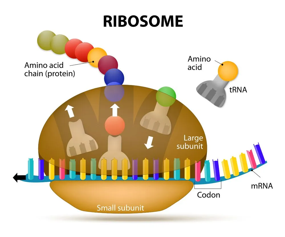
Before we uncover how multiple codons can code for a single amino acid, let’s learn a bit more about the players involved.

mRNA: The Starting Point Of Translation
Messenger RNA or mRNA is a single-stranded piece of RNA. It is synthesized during a process called transcription, where the information from the DNA is copied or transcribed into an mRNA strand. The double-stranded DNA helix opens up and an mRNA strand identical to the 5’ to 3’ coding strand is produced. Note that ribonucleic acid (RNA) contains the nucleotide uridine (U) in place of thymine (T), which is the only difference between the mRNA and the coding (5’ to 3’) strand of the DNA.
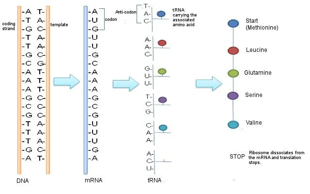
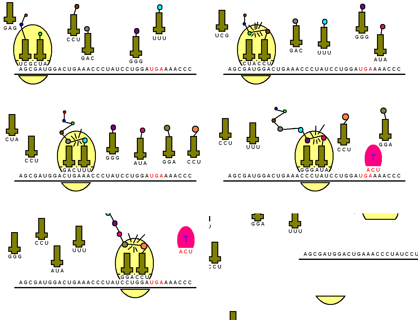
The mRNA then makes its way to the protein-making machinery, the ribosomes. The ribosomes read the information in mRNA to make proteins. This is called translation, like translating instructions to make something.
The information in the mRNA is read three nucleotides at a time in sections called codons. Each codon specifies a certain amino acid.
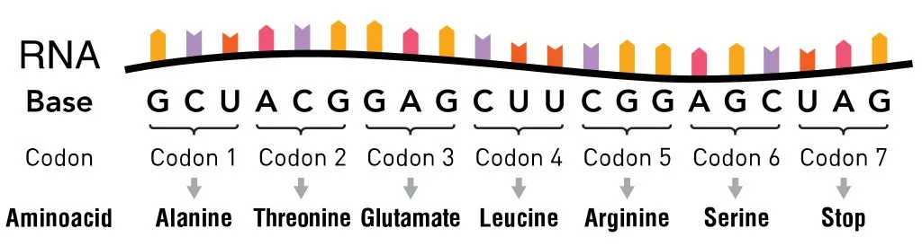
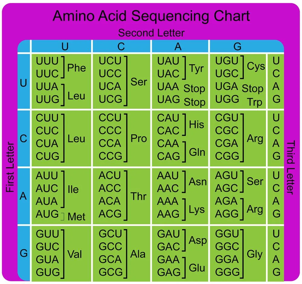
For example, the codon AUG codes for the amino acid methionine (Met). AUG also codes where the protein information starts, called the start codon, which is required to initiate the translation process. Thus, methionine is always the first amino acid in an amino acid chain. The amino acid tryptophan (Trp) is indicated by the codon UGG.
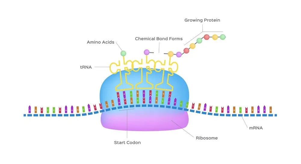
Codons don’t only code for amino acids; they also code for when to terminate the translation process. They are collectively known as stop codons and are UAG, UAA and UGA.
Hence, a codon is like a three-letter password that is required to obtain an amino acid and marks where the instructions to start and stop making the protein are found.
tRNA: The Assembly Point Of Translation
The transfer RNA or tRNA is also single-stranded, but is folded, unlike the straight mRNA. It serves as a physical link between the mRNA and the amino acids by interpreting the mRNA and transferring the right amino acids to their place while making the protein. If the translation machinery was a factory, the tRNA would be the factory workers who interpret the instruction manual (the mRNA) to carefully put products in the specified order.
The tRNA is structured in such a way that it has an anticodon loop on one end and an acceptor arm/stem on the opposite end.
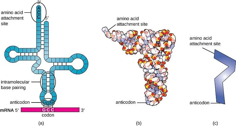
As the name suggests, the anticodon loop is complementary and antiparallel (3’ to 5’) to the mRNA codons. That is, the tRNA consists of the 3 nucleotides that bind to the ones present on the mRNA. Thus, guanine (G) and cytosine (C) bind to each other and adenine (A) and uridine (U) bind to one another.
So, considering the codon for methionine, 5′-AUG-3′, the matching tRNA carries the anticodon 3′-UAC-5′ (written in the standard 5′-to-3′ direction, that's CAU). Each anticodon base pairs with its complement on the codon: A–U, U–A, G–C.
This type of pairing is known as Watson-Crick base pairing. The pairing is like the attraction between highly specific magnets. Anti-codons on the tRNA ensure that it lines up against the correct codon on the mRNA. Furthermore, as codons are read in the 5’ to 3’ direction, anticodons present on the tRNA are positioned in the 3’ to 5’ direction.
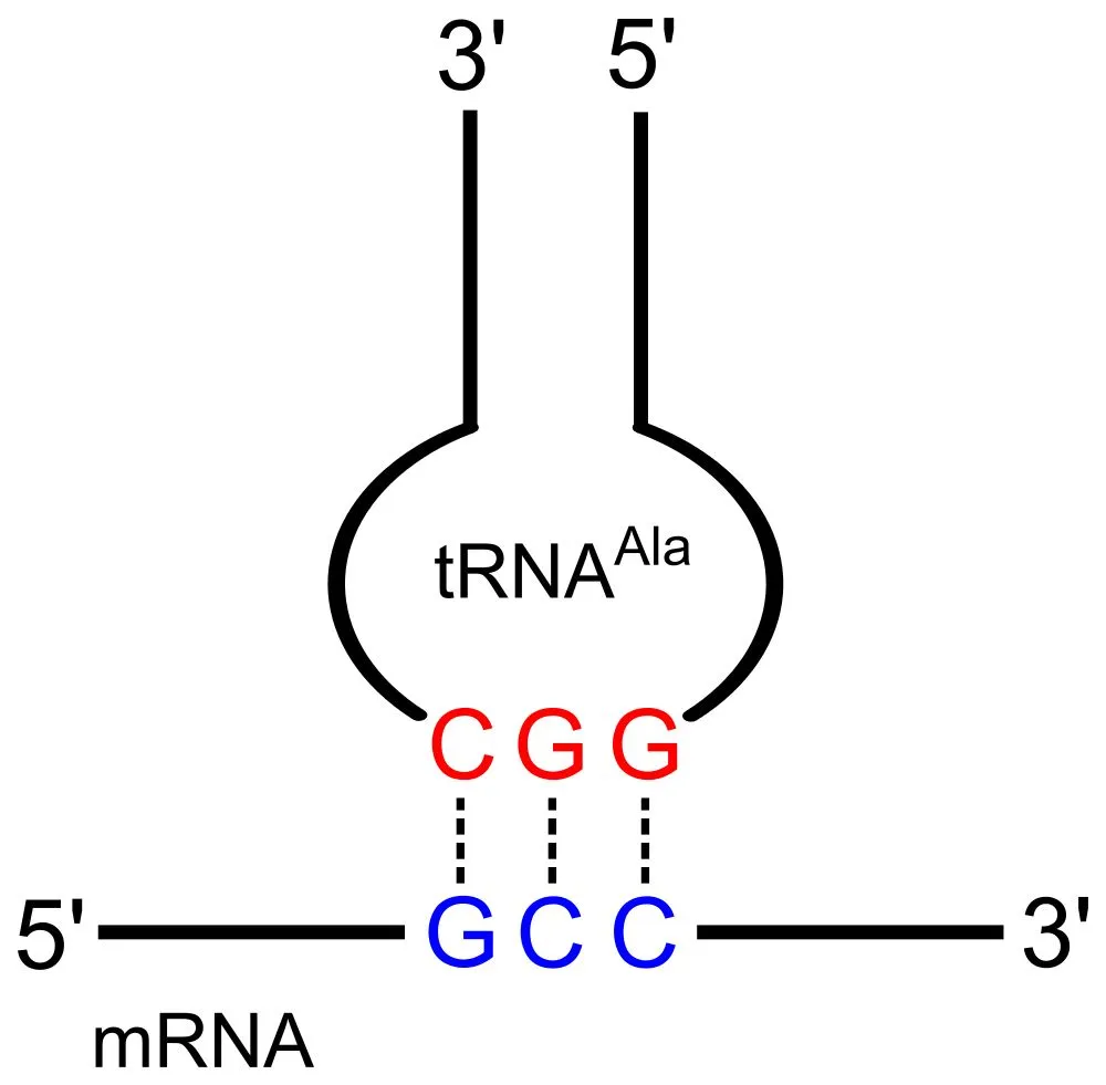
This means that the 1st codon base binds to the 3rd anticodon base and so on. Note that each amino acid has its very own tRNA, which correctly positions it in the polypeptide chain due to the base-pairing between the codon and anticodon.
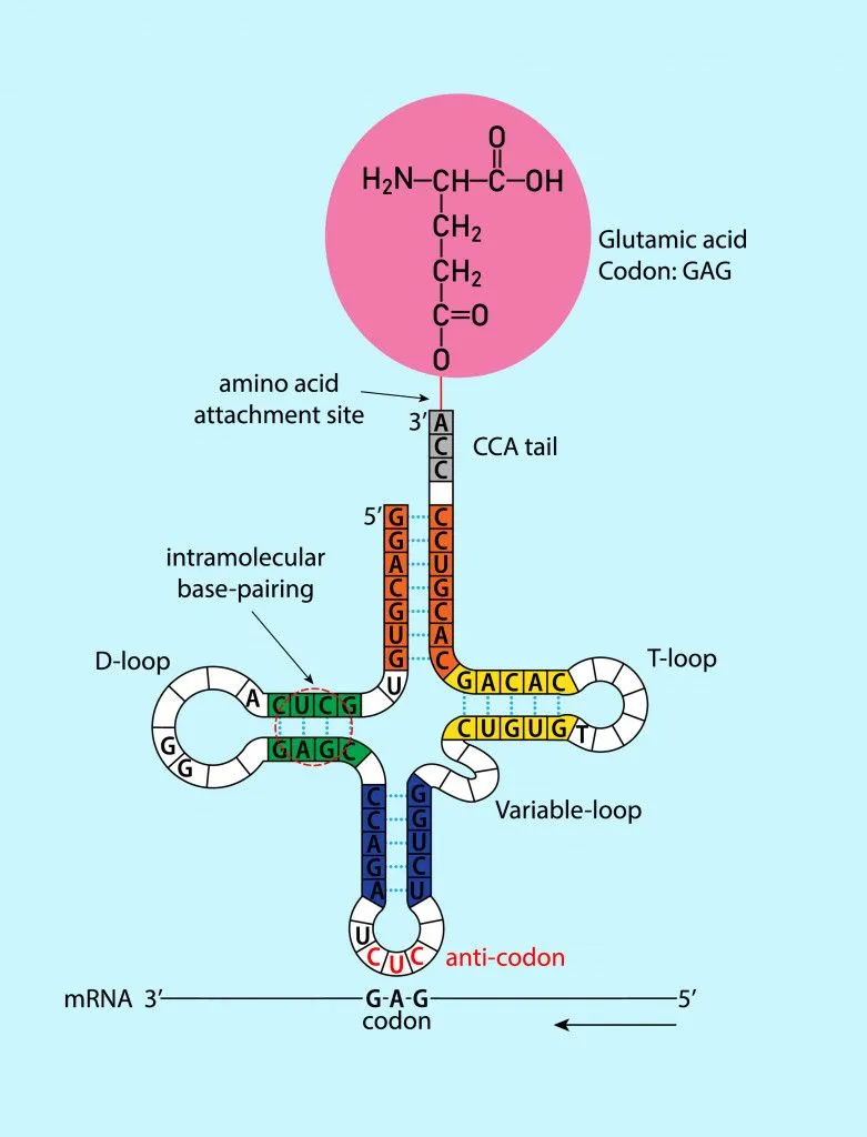
However, like many things in biology, this is also a little more complicated. Each amino acid can be specified by more than one codon. Additionally, the base-pairing rules between the codon and anticodon are not equally binding for all bases. We will learn more about this peculiar phenomenon next!
Wobble Hypothesis: Why 20 Amino Acids Have 64 Codons?
As we know, DNA is made up of 4 nucleotide bases (A, G, T, C). These letters (bases) are read three at a time, which means that there are 64 (4 x 4 x 4) possible combinations of these triplets or codons.
Of the 64 codons, 3 are stop codons, which we mentioned previously. These three stop codons do not code for amino acids and only terminate the process of translation. Therefore, we are left with 61 codons for just 20 amino acids.
The only logical option is that a single amino acid can be coded by multiple codons. But, in theory, 61 different tRNAs would be required to read every different sense codon, yet this isn't the case, as cells actually get by with fewer than 50 distinct tRNA anticodons.
This observation implies that there might be some leeway between codons and anticodons matching up. Consider the amino acid valine, which is coded by 4 codons: GUU, GUC, GUA, GUG. Notice that only the third base changes, while the first two are the same.
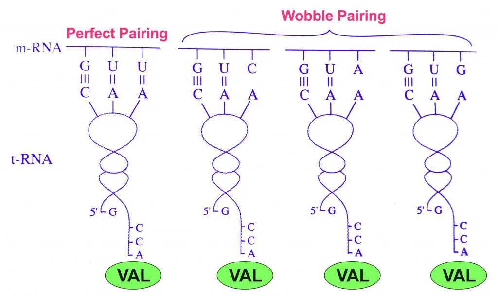
This variance observed only on the 3rd base of the codon led to the ‘Wobble Hypothesis’, proposed by Francis Crick (one of the discoverers of DNA's double-helix structure) in 1966.
The Wobble Hypothesis explained this variance by revealing that the rules of Watson-Crick base-pairing were not followed at the last position of the codon. That is, the bond between the 3rd letter (nucleotide base) and the 1st letter of the anticodon may show unusual bonding (non A-T/ non G-C). Hence, it was allowed to ‘wobble’.
This meant that while the 1st and 2nd bases of the codons strictly adhered to the rules of Watson-Crick base pairing by binding to the 3rd and 2nd base of the anticodon, respectively, non-Watson-Crick base pairing was allowed between the 3rd base of the codon and 1st base of the anticodon. Hence, some tRNAs can recognize many different codons. It also explains the pattern of redundancy in the genetic code (many codons for a single amino acid).
The rules of the Wobble Hypothesis are as follows:
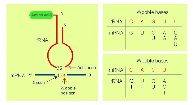
- The first two bases of the codon and the last two bases of the anticodon undergo normal Watson-Crick base pairing. That is, hydrogen bonds are formed between adenine (A) and uridine (U), guanine (G) and cytosine (C), only.
- Less stringent rules of base pairing apply at the remaining position and non-Watson-Crick base pairing may take place. The bases that undergo such pairing are also referred to as wobble base pairs. This allows the anticodon of a single form (amino acid specific) of tRNA to pair with more than one codon in the mRNA.
- When uridine (U) is present in the remaining (1st base of anticodon) position, it can recognize adenine (A) or guanine (G) only.
- When guanine (G) is present in the remaining (1st base of anticodon) position, it can recognize uridine (U) or cytosine (C) only.
- If the modified base inosine (I) is present in the remaining (1st base of anticodon) position, it can recognize uridine (U), cytosine (C) or adenine (A).
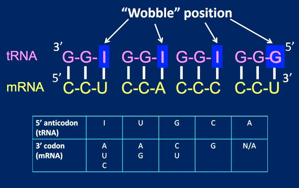
The reason for the wobble base pairing is a slip-up on the part of the ribosome. The ribosome has mechanisms in place to check whether the 1st and 2nd codon bases are complementary to the 3rd and 2nd anticodon bases, respectively. However, the ribosome doesn’t have a mechanism to check whether the 3rd codon base on the mRNA and 1st anticodon base on the tRNA match.
How Many Codons Code For One Amino Acid?
This is where a lot of the confusion creeps in, so let's clear it up. A single amino acid is specified by one codon at a time, and a codon is always a set of three mRNA bases. So it takes three bases (not three codons) to spell out one amino acid. What degeneracy adds is that the same amino acid can be written using several different three-letter codons.

How many codons map to a given amino acid varies from one to the next. Of the 64 codons, 61 are sense codons that specify amino acids, while 3 are stop codons. Spread those 61 across just 20 amino acids and the share works out unevenly:
- Six codons each: leucine, serine and arginine, the most flexible of the set.
- Four codons each: valine, alanine, glycine, proline and threonine.
- Three codons: isoleucine.
- Two codons each: phenylalanine, tyrosine, histidine, glutamine, asparagine, lysine, aspartate, glutamate and cysteine, among others.
- One codon each: methionine (AUG) and tryptophan (UGG), the only two amino acids with a single, exclusive codon.
So if you are ever asked to name an amino acid that has more than one codon, leucine is a safe answer, since it has six. And if the question flips to which amino acids have just one, the pair to remember is methionine and tryptophan.
How Do You Read The Genetic Code To Find An Amino Acid Sequence?
Once you have an mRNA sequence, working out the protein it codes for is mostly a matter of looking up each codon in the table above. The trick is to read in the right direction and keep to the right frame.
Start reading the mRNA from its 5′ end towards its 3′ end, the same way the ribosome travels. Find the first AUG (the start codon), then break everything after it into non-overlapping groups of three. That grouping is the reading frame, and shifting it by even one base reshuffles every codon downstream.
Let's translate a short strand, 5′-AUG GCC UCA GGG UGA-3′:
- AUG → methionine (and the signal to begin)
- GCC → alanine
- UCA → serine
- GGG → glycine
- UGA → stop
Read off in order, that gives the peptide methionine-alanine-serine-glycine, and the UGA stop codon tells the ribosome to release the finished chain. Because UGA, UAA and UAG do not stand for any amino acid, they never appear in the protein itself; they simply mark the end of the message.
The Significance Of The Wobble Hypothesis
Slip-up or not, because of the wobble base, there is a lesser chance of mistakes during translation. For example, if the leucine (Leu) codon CUU was misread as CUA, CUG or CUC during transcription, the codon would still be translated as leucine (Leu) during protein synthesis.
Furthermore, protein synthesis is also safeguarded against mutations of a single base. Thus, if the 3rd base in the valine codon GUU mutates and changes to GUC, GUA or GUG, it will still be translated correctly.
Wobble and the degeneracy of the genetic code also reduce the number of tRNAs required by a cell. For instance, for the four different codons of glycine, there are only 3 tRNAs present in E.coli.
So, while it is true that multiple codons can code for a single amino acid, it is the ‘wobble’ observed between the 3rd codon base and the 1st anticodon base that makes this flexibility possible.
What's The Advantage Of Having Multiple Codons?
Beyond trimming the number of tRNAs a cell has to build, degeneracy hands the genome a built-in safety net against mutations. DNA is copied countless times, and now and then a base gets swapped by mistake. When that single-base swap, a point mutation, lands on the third position of a codon, the redundancy of the code often means the codon still spells out the same amino acid.
A change that leaves the amino acid untouched is called a silent (or synonymous) mutation, because the finished protein is identical. For example, the codon GAA codes for glutamate; if its last base flips so that it reads GAG, the ribosome still inserts glutamate and the protein is none the wiser. Contrast that with a missense mutation, where the swap does change the amino acid (the change behind sickle-cell disease), or a nonsense mutation, where a codon turns into a premature stop and cuts the protein short. By absorbing so many third-position changes as silent ones, the degenerate code keeps a large share of random mutations from ever reaching the protein.
That said, "silent" is not always perfectly silent. Cells tend to favor some synonymous codons over others, so a swap to a rarely used codon can subtly slow translation or alter how the mRNA folds. The amino acid sequence stays the same, but the finer details of gene expression can still shift.
References (click to expand)
- Crick, F. H. C. (1966, August). Codon—anticodon pairing: The wobble hypothesis. Journal of Molecular Biology. Elsevier BV.
- (2014) Intronic and plasmid-derived regions contribute to the large .... National Center for Biotechnology Information
- BB331 Lecture 12 - oregonstate.edu
- How the ribosome decodes a modified 'wobble' position .... The European Synchrotron Radiation Facility
- Wobble Hypothesis - Newcastle University. Newcastle University
- Chapter 5. Genetic Code, Translation, Splicing - Biology. Kenyon College
- Amino Acids Are Encoded by Groups of Three Bases Starting from a Fixed Point - Biochemistry - NCBI Bookshelf - www.ncbi.nlm.nih.gov
- Hoerter, J. E., Ellis, S. R. Biochemistry, Protein Synthesis. StatPearls. NCBI Bookshelf.
- Popovic, A., Orrick, J. A. Biochemistry, Mutation. StatPearls. NCBI Bookshelf.
- Genetic Code: Codon Assignments and Degeneracy. Biology LibreTexts.