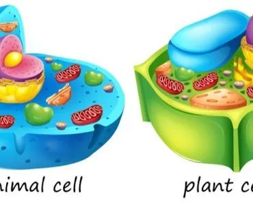
Endocytosis is the umbrella process by which a cell engulfs material, wrapping it in a membrane pocket that pinches off into a vesicle. Phagocytosis ("cell eating") takes in large particles or whole cells; pinocytosis ("cell drinking") takes in fluid and dissolved molecules. Both are forms of endocytosis, and all of them are active processes that require the cell to spend energy.
The human body is packed with more mysteries than we will ever understand, but we do understand the crucial processes that allow each of our cells, organs, tissues and muscles to function. The microscopic movement of material between a cell and its surroundings is an incredibly important process for human beings, and it relies on a family of related mechanisms, including endocytosis, phagocytosis and pinocytosis.
What Is Endocytosis?
Have you ever watched bubbles floating on top of water? When they collide, the smaller bubbles are often swallowed up by the larger ones. On an even smaller scale, at the cellular level, the cells in our body can do something similar, taking up molecules from the extracellular fluid. This process is called endocytosis, and is currently happening in millions of your cells at this very instant!
While it may seem strange to think of a cell needing to consume something, there are many reasons that a cell requires processes like endocytosis, such as absorbing essential nutrients from the extracellular fluid, consuming entire microorganisms, or transporting dead or dying cells to where they can be eliminated.
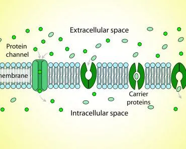
The actual process of endocytosis is simple and straightforward, although there are a number of different forms. Basically, once a molecule or other substance is encountered by the cell, the cell’s plasma membrane folds inwards surrounding the material, forming a pocket. Eventually, that pocket of membrane closes, and a vesicle is formed, which can separate from the membrane in the inside of the cell and then move to the organelle where it can be used.
Endocytosis is the opposite process to exocytosis, which is the means by which a cell can export molecules, whether they are proteins, neurotransmitters, waste products or any other cellular material. We’ve covered all the details of exocytosis in this article.
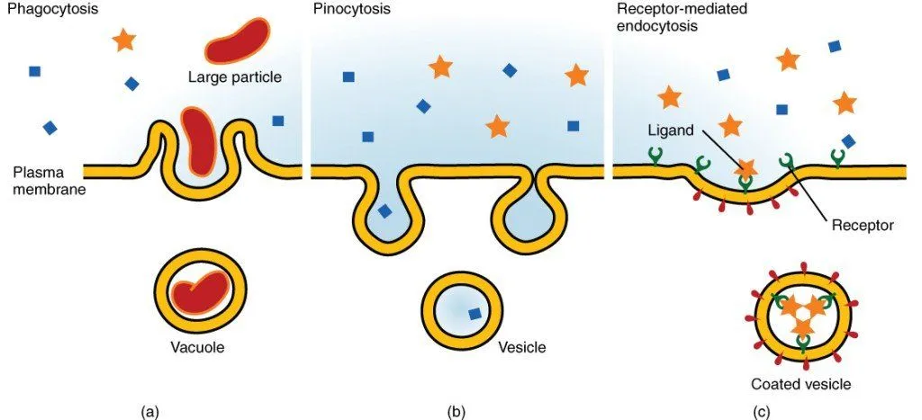
There are two main types of endocytosis, phagocytosis and pinocytosis, which are defined by the kind of material the cell takes up. Beyond these, there are more selective routes that we will also briefly discuss: receptor-mediated endocytosis (which is usually clathrin-mediated) and the clathrin-independent caveolae pathway.
Phagocytosis
When certain types of cells (phagocytes) consume an entire other cell or macromolecule, this form of endocytosis is called phagocytosis. This process can occur in various cells, including amoeba and white blood cells. For example, when a white blood cell is sent to the site of inflammation or infection, it can then consume unwanted bacteria, viruses, foreign particles or even pieces of dust. At that point, the unwanted material can be broken down into simple, usable molecules, or discarded from the body as waste.
Depending on the size of the material being consumed, the speed of this process may vary. Since the molecules being consumed are typically quite large, the vesicles that must be formed require a moderate amount of energy and resources, in the form of ATP. Similar to the process of exocytosis, this is a form of active transport, as the molecules cannot passively be consumed through the cell membrane. It is important to remember that only certain specialized cells are able to perform phagocytosis, whereas pinocytosis, explained below, is something that all cells must perform.
Pinocytosis
The second main form of endocytosis is pinocytosis, by which a cell takes in a pocket of extracellular fluid and micro molecules at random. This process is also informally called “cell drinking”, which is an accurate description of this fluid uptake. Similar to phagocytosis, the plasma membrane of the cell extends to form a pocket or a bud, and then closes the pocket. This forms a vesicle within the cell, which is then pinched off and can be moved to other areas of the cell in the cytosol. This form of endocytosis is also important because it can transport extracellular liquid through the cell without it interacting or affecting the cytoplasm. As mentioned, this cell drinking occurs in every type of cell. And yes, like phagocytosis, pinocytosis is active transport, so it too runs on energy in the form of ATP; reshaping the plasma membrane to pinch off a vesicle simply doesn't happen for free.
Receptor-Mediated Endocytosis: Clathrin-Mediated And Caveolae-Mediated
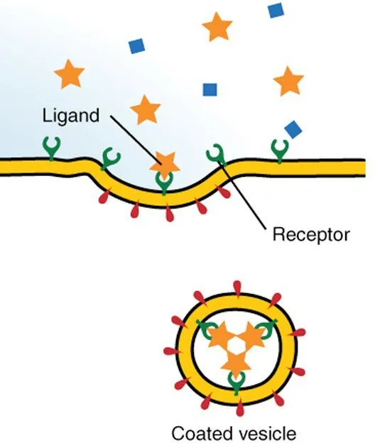
The last two types of endocytosis are slightly more specialized, and require different cellular technology to occur. In clathrin-mediated endocytosis, larger macromolecules are taken into a cell through concentrated areas of clathrin-coated vesicles. Clathrin is a coat protein that assembles into a basket-like cage around these pits. These pits on a plasma membrane facilitate the easier movement of large molecules, and are found on almost all cell types. Certain molecules have different receptors, and the clathrin-coated pits are able to provide the necessary “keys” for the membrane “lock”.
Caveolae-mediated endocytosis is similar to clathrin-mediated endocytosis, except the vesicles are not coated in clathrin but in another membrane protein called caveolin. They are small, flask-shaped pits waiting below the cell surface, and they serve a function similar to the clathrin-coated pits. Caveolae are extremely abundant in certain cell types, such as fat cells (adipocytes) and the endothelial cells lining blood vessels, where they can occupy a large fraction of the plasma membrane surface (commonly cited at upwards of 20%, and even more in some adipocytes).
What's The Difference Between Phagocytosis And Pinocytosis?
First, let's clear up a common mix-up. Is phagocytosis the same thing as endocytosis? Not quite. Endocytosis is the umbrella term for any process in which a cell pulls material inside by wrapping it in a piece of its own membrane. Phagocytosis is one specific type of endocytosis, and pinocytosis is another. In other words, every act of phagocytosis is endocytosis, but not every act of endocytosis is phagocytosis, in the same way that every poodle is a dog, but not every dog is a poodle.

Phagocytosis was the first of the two to be noticed. The zoologist Élie Metchnikoff (also spelled Ilya Mechnikov) made his original observations in the 1880s while studying marine invertebrates, and his work on these hungry cells helped earn him the Nobel Prize in 1908. Macropinocytosis, the "big gulp" form of pinocytosis, was described in mammalian cells in 1931 by Warren Lewis, who showed that macrophages and cancer cells ruffle their membranes and swallow the surrounding fluid, and captured the process in time-lapse films.
So how do the two actually differ? It comes down to what the cell is taking in, and what it plans to do with it.
| Feature | Phagocytosis ("cell eating") | Pinocytosis ("cell drinking") |
| What goes in | Solid particles larger than about 0.5 micrometers (µm), such as bacteria, dead cells and cellular debris | Extracellular fluid and whatever happens to be dissolved in it |
| Selectivity | Selective: receptors on the cell must first recognize the target | Largely non-selective: the cell takes a gulp of whatever is around it |
| Who does it | Mainly specialized immune cells, such as neutrophils and macrophages | Many cell types, from immune cells to the endothelial cells that line blood vessels |
| How the membrane moves | Actin-driven "arms" called pseudopodia reach out and wrap around the particle | The membrane folds or ruffles inward to trap a pocket of fluid |
| Vesicle size | Big enough to hold the particle it swallowed (a phagosome) | Macropinosomes are 0.2 to 5 µm across; other forms make smaller vesicles |
| What happens next | The phagosome fuses with lysosomes to become a phagolysosome, where the cargo is destroyed | The vesicle shrinks as it is processed, and much of its membrane is returned to the cell surface |
The fate of the cargo is perhaps the biggest difference. Phagocytosis has even been described as a "destructive" form of endocytosis. Once a particle is sealed inside its phagosome, that compartment matures in stages, fusing with lysosomes and growing steadily more acidic, until it reaches a pH as low as 4.5. Few bacteria enjoy that kind of hospitality. Pinocytosis, by contrast, is less a hunt and more a sip: the cell isn't chasing any particular target, so it samples the surrounding fluid, dissolved nutrients and all.
Is Endocytosis Active Or Passive Transport?
This is one of the most common questions about endocytosis, and the short answer is: active. Both phagocytosis and pinocytosis are active processes. The reason, however, is more interesting than a one-word answer.
In passive transport, substances simply drift from an area of higher concentration to an area of lower concentration, and the cell doesn't have to spend any of its energy to make that happen. Diffusion is the classic example. Endocytosis doesn't work like that. The cargo isn't slipping through the membrane at all; instead, the cell physically reshapes its own membrane to package the cargo, which is why endocytosis is grouped under bulk transport. Phagocytosis, pinocytosis and receptor-mediated endocytosis are all classified as active transport, because the cell must spend energy to carry them out.
So where does that energy go? Several key steps depend on energy-consuming molecular machinery:
- Cutting the vesicle loose: A protein called dynamin assembles into a helical collar around the narrow neck of a budding vesicle. By hydrolyzing GTP (guanosine triphosphate), dynamin changes shape and triggers fission, snipping the vesicle free from the membrane.
- Stripping off the coat: Once a clathrin-coated vesicle is inside the cell, its clathrin cage has to come off. That job falls to a chaperone protein called Hsc70, long known as the "uncoating ATPase", which uses ATP to pull the coat apart so the clathrin can be used again.
- Acidifying the compartment: After phagocytosis, proton pumps called V-ATPases use ATP to push hydrogen ions into the phagosome, making it acidic enough to break down what was swallowed.

What happens when one of these machines breaks down? Fruit flies offered an early clue. Flies carrying a mutation called shibire, first identified in 1973, are temperature-sensitive: under certain conditions, they become paralyzed. Scientists later traced the mutation to the gene for dynamin. The paralysis happens because the flies' nerve cells run out of synaptic vesicles, the tiny packets that store and release neurotransmitters. Normally, neurons recycle those vesicles through endocytosis, but without working dynamin, the endocytic pits get stuck at the membrane with "collars" around their necks, unable to pinch off. The nerve signals stall, and so does the fly.
What Are Some Real-World Examples Of Endocytosis In The Human Body?
Endocytosis might sound like an abstract textbook topic, but it's happening throughout your body right now. Here are a few places where it really matters.
Cleaning Up Invaders And Dead Cells
When microorganisms invade the body, white blood cells called neutrophils remove them by phagocytosis. Just as important, if less dramatic, is the daily housekeeping. Phagocytes such as macrophages help clear away the billions of cells that are turned over in the body every day. How do they know which cells to eat? Dying cells display a molecule called phosphatidylserine on their surface, which healthy cells don't normally show, and its levels can rise as much as 300-fold. Healthy cells, meanwhile, carry a protein called CD47, which binds a receptor on the phagocyte and delivers an inhibitory signal that blocks the actin assembly needed for engulfment. It's essentially a molecular "don't eat me" badge.
Delivering Cholesterol
Cholesterol is a structural component of your cell membranes, and most of the cholesterol in your bloodstream travels inside particles called low-density lipoprotein (LDL), each carrying a core of roughly 1,500 cholesteryl ester molecules. Cells capture LDL through receptor-mediated endocytosis: LDL binds to LDL receptors sitting in clathrin-coated pits, the pits pinch off as coated vesicles, and the cholesterol is released inside the cell while the receptor is recycled back to the surface. Michael Brown and Joseph Goldstein shared the 1985 Nobel Prize in Physiology or Medicine for these discoveries.

Their key clue came from familial hypercholesterolemia (FH), an inherited disorder in which functional LDL receptors are partly or completely missing. Brown and Goldstein found that cells from patients with the most severe form of FH completely lacked functional LDL receptors, so LDL stayed in the blood instead of being pulled into cells, where it could accumulate in artery walls. Today, the CDC estimates that FH affects about 1 in 311 people. It's a sobering reminder that one faulty doorway can raise a person's risk of heart attack.
Letting Viruses In (Unintentionally)
Not every visitor at the door is welcome. Many endocytic pathways are exploited by viruses, bacteria and toxins to gain entry into cells. HIV-1, for example, can be taken into macrophages inside large vesicles that resemble macropinosomes. Researchers are now trying to turn the tables by using these same routes to deliver therapeutic drugs into cells.
Feeding Tumors
Some cancer cells have learned to drink their way to growth. Pancreatic cancer cells with mutations in the Ras gene can use macropinocytosis to gulp down proteins from their surroundings and break them down for amino acids, essentially reverting to the feeding habits of amoebae. It's an unsettling example of how a basic cellular skill can be turned to less friendly ends.
A Final Word
These processes are essential to our survival every day, and yet we have no conscious control over them. They are yet another example of how impressive and intricate our bodies are, and how even the simplest processes at the smallest level of life can have measurable impacts on health. As is so often seen and commented on in science, the microcosm reflects the macrocosm. Just as we consume food from our surroundings, so too do our cells, and while we don’t capture our hamburgers with a plasma membrane, there are certainly parallels if you pay attention!
References (click to expand)
- Endocytosis | Drubin/Barnes Lab.
- Molecular Expressions Cell Biology: Endosomes.
- Endocytosis. The Cell: A Molecular Approach. NCBI Bookshelf.
- Endocytosis - academic.brooklyn.cuny.edu:80
- ENDOCYTOSIS - classes.kumc.edu
- Phagocytosis. StatPearls. NCBI Bookshelf.
- Phagocytosis: A Fundamental Process in Immunity. BioMed Research International (2017).
- 5.4 Bulk Transport. Biology 2e. OpenStax.
- 5.2 Passive Transport. Biology 2e. OpenStax.
- Macropinocytosis in Different Cell Types: Similarities and Differences. Membranes (2020).
- The origins and evolution of macropinocytosis. Philosophical Transactions of the Royal Society B (2019).
- Dynamin, a membrane-remodelling GTPase. Nature Reviews Molecular Cell Biology (2012).
- The role of molecular chaperones in clathrin mediated vesicular trafficking. Frontiers in Molecular Biosciences (2015).
- Endocytosis: Past, Present, and Future. Cold Spring Harbor Perspectives in Biology (2014).
- In situ exploration of characteristics of macropinocytosis and size range of internalized substances in cells by 3D-structured illumination microscopy. International Journal of Nanomedicine (2018).
- The Nobel Prize in Physiology or Medicine 1985: Press release. NobelPrize.org.
- About Familial Hypercholesterolemia. Centers for Disease Control and Prevention (CDC).