The pupillary light reflex is a reflex that controls the diameter of the pupil when it is exposed to varying intensities of light. This allows the eyes to adjust in response to bright or dim lights.
Walk into any room and switch on the light; everything seems perfectly in its place. Now, switch off the light and try to see what is in the room. Give yourself a bit of time and you’ll be amazed at how clearly you can still identify various items in the space. Although you may not be able to see them as perfectly as before, you can still see well enough so you won’t trip in the dark!
The manner in which your eyes are able to adapt to the lighting and allow you to see in the dark is fascinating! The eye is a beautiful part of the body that helps us see marvelous images from the world around us. The pupil is able to adjust to light in the surroundings, allowing us to see in both light and dark environments. The pupillary light reflex is the reflex that controls the diameter of the pupil when it is exposed to varying intensities of light.
How Does The Eye Work?
Vision is a complex process that involves the coordinated and simultaneous activity of the brain and the eye. The light entering the eye gets converted to an electrical response called a nerve impulse. These nerve impulses travel to the brain along the optic nerve in order to create the final image. The optic nerve is the nerve that transmits visual information from the eye to the brain.
To better understand how light travels from the retina to the brain, let’s take a closer look at the eye!
The most important parts of the eye include the iris, the cornea, the lens and the retina. Each of these plays an indispensable role for us to see a clear image. The eye or the eyeball is located in the eye socket, but we can only see the front part of the eye.
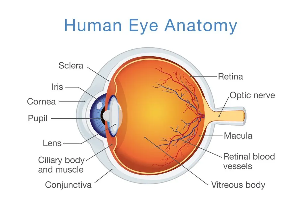
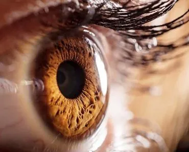
The white part of the eye is the sclera, which is the visible section of the outer layer of the eyeball. The colored part of the eye is the iris, which has a small disc-like shape with a hole in it called the pupil. The pupil is black, as almost no light can escape from it. It is the pupil that either contracts or dilates, depending on the amount of light that reaches it. A transparent layer called the cornea covers the iris and the pupil. The cornea is like a dome around the iris and behind the cornea is a fluid called the aqueous humor, which helps to cleanse the eye and provide the required nutrients to the eye. The cornea also protects the eye from foreign particles and injury. Our eyelids and eyelashes serve the same function, although many people (myself included) probably wish there was something to prevent eyelashes from entering the eye!
The eye has 2 other very important components, the lens and the retina. The lens is attached to muscles with the help of strong fibers. The contraction of these muscles will change the shape of the lens. Light rays that pass through the pupil reach the lens located behind it. The role of the lens is to focus the light on the retina. The path of light that enters the eye changes (refracts) to different extents, depending on the shape of the object. Refraction occurs when light enters different mediums. In the eye, light travels from air into a liquid-like medium of the cornea, which causes a change in its pathway. The process of change in the pathway of light is called accommodation and it allows the eye to focus on objects located either close by or far away.
The retina is the innermost tissue of the eye and is a light-sensitive portion of our visual system. The retina contains millions of sensory cells, including light-sensing cells called photoreceptor cells, which are designated as either rods or cones. Rods function in dim light and provide the contrast of black-and-white vision, whereas cones function in well-lit environments and are able to perceive different colors.
The refraction caused by the lens creates a sharp image on the retina. The sensory cells present on the retina receive these light signals and transmit them to the brain via the optic nerve.
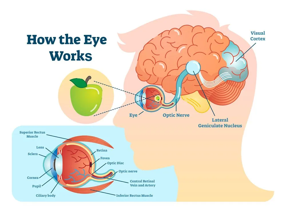
The main role of the optic nerve is to transfer visual information from the retina to the vision centers of the brain with the help of electrical impulses. The optic nerve is made up of nerve cells and is a critical part of the central nervous system.
In simple terms, the nerve signals from the rods and cones are sent to the brain via the optic nerve. Inside the brain, these signals are converted to create the images that we see.
How Does The Eye Respond To Light?
Up to this point, we’ve discussed how light enters the eye and travels to the brain via the optic nerve to create the final image. Now, let’s further discuss how the eye responds to different intensities of light, which is closely related to the processes explained above.
I recently went to my best friend’s wedding and seeing the newly married couple made my heart fill with joy. At that moment, I took two pictures, one with the help of a digital camera, while the other was a “mental picture”. Basically, I focused my gaze on a scene, in the hopes that my brain would store that particular image, just like on a regular memory card. Interestingly, the eye functions much like a camera, adjusting the light that enters the pupil so that we get a perfect image.
When the light entering the eye is too bright, the pupil constricts or shrinks, allowing less light to enter the eye. When you point a bright light directly at your eye, you can observe how swiftly the pupil constricts, almost instantaneously. The pupils constrict as a way of protecting the retina from dangerously bright lights.
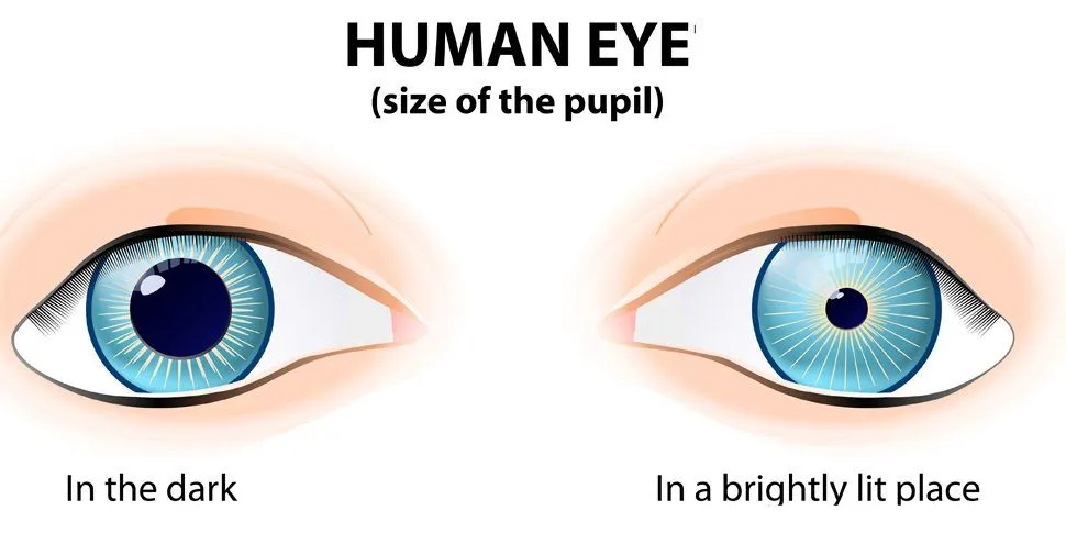
At the other extreme, when you enter a dimly lit area, the pupil dilates or expands, allowing for more light to enter the eye. In fact, dilated pupils are often seen as a sign of beauty, which is one of the reasons why candle-lit dinners are often linked with romance!
What Is The Pupillary Light Reflex Pathway?
So far we have seen what the pupil does, but not how the message actually gets there. The beauty of the pupillary light reflex is that it is a true reflex; it happens automatically, without you having to think about it, much like snatching your hand away from a hot stove. The whole loop is called a reflex arc, and it has two halves: an incoming (afferent) limb that carries the “there is light” signal to the brain, and an outgoing (efferent) limb that carries the “shrink the pupil” instruction back to the eye.

The incoming half begins at the retina. When light lands on it, the signal travels along the optic nerve (also known as cranial nerve II) through the optic chiasm and optic tract to a small relay station in the midbrain called the pretectal nucleus. Here is the clever bit: each pretectal nucleus connects to both sides of the brain, passing the signal on to two clusters of cells called the Edinger-Westphal nuclei, one linked to each eye.
From the Edinger-Westphal nuclei, the outgoing half takes over. These parasympathetic fibers ride along the oculomotor nerve (cranial nerve III) to a small hub behind the eye called the ciliary ganglion. From there, short nerves reach the sphincter pupillae, a ring of muscle in the iris that tightens like a drawstring to shrink the pupil. When you step into the dark instead, a separate set of sympathetic nerves activates the dilator pupillae muscle, which pulls the pupil wide open. In short, the parasympathetic nervous system closes the pupil, while the sympathetic nervous system opens it.
Why Do Both Pupils React When A Doctor Shines A Light In One Eye?
Have you ever noticed a doctor shining a small torch into one eye and then the other during a check-up? They are testing this very reflex. Because each pretectal nucleus wires into both Edinger-Westphal nuclei, light shone into a single eye makes both pupils constrict at the same time. The lit eye’s pupil shrinking is called the direct response, while the other pupil shrinking, even though no light entered it, is called the consensual response.
Doctors put this to work with a simple check called the swinging flashlight test. In a dimly lit room, the examiner swings a light back and forth between the two eyes and watches how the pupils react. In a healthy person, both pupils stay neatly constricted no matter which eye the light is on. If the light swings onto one eye and both pupils surprisingly widen instead of staying small, it suggests that eye is sensing less light than the other, a sign known as a relative afferent pupillary defect, or Marcus Gunn pupil (named after the ophthalmologist Robert Marcus Gunn).
This matters because the pupils are a window onto the nerves behind them. A weak or unequal light reflex can be an early clue to trouble along the visual pathway, such as optic neuritis, glaucoma, a retinal detachment, or damage to the optic nerve. It is also why checking the pupils is one of the first things a doctor or paramedic does after a head injury; a quick, symmetric reflex is a reassuring sign that the reflex arc, and the brain regions it passes through, are still doing their job.
How Are We Able To See In The Dark?
The main players in vision are the rods and cones. The rods are extremely efficient in dim light, as even a small amount of light is able to trigger them. They are able to accurately detect light, contrast and movement, but they’re unable to identify color.
The pupil dilates as a result of low light and allows as much light as possible to enter the eye. This light then reaches the retina and the rods convert the light energy to electrical energy, with the help of the optic nerve, to produce the final image. There’s one major difference; given that the rods are unable to identify color, the image seen in low or dim light is always in black and white.
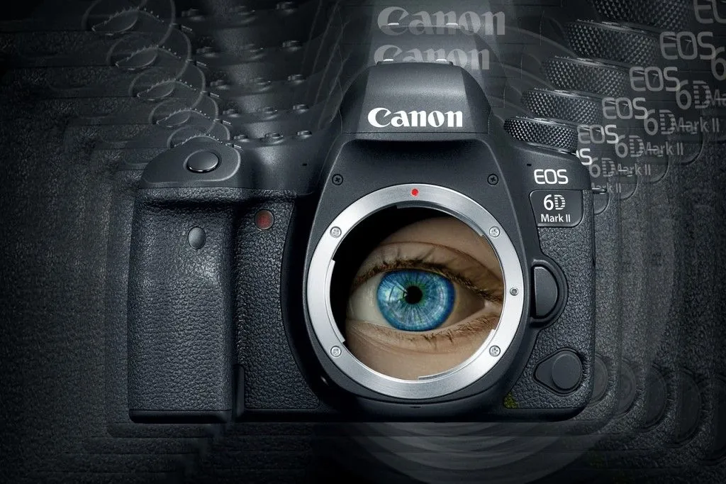
The pupillary light reflex is a very unique and interesting way that the eye captures images and manages light exposure. Almost like a camera, the eye adjusts the amount of light that enters to create the perfect setting; this leads to the formation of an image by the brain. However, the next time you take a picture with a camera, remember to manually adjust the aperture for perfect lighting, as the pupillary reflex is a specialty of the eye alone!
Why Is It So Hard To Judge High Catches Under Floodlights?
Anyone who has watched a night cricket or baseball game has seen it happen: a fielder settles under a towering skier, only to spill a catch that would have been routine in daylight. Part of the reason lies in the very light-and-eye relationship we have been exploring.

Judging where a high ball will land depends on depth perception. Up close, your two eyes see slightly different views of an object and your brain fuses them to sense distance, a trick called stereopsis. This works beautifully for nearby things, but it grows weaker with distance, because for a ball tens of meters up in the air the two eyes see almost exactly the same picture. So your brain instead leans on monocular cues, the clues a single eye can use, such as the ball’s changing size and its position against the background.
This is exactly where a dark night sky lets you down. Against a plain, textureless black backdrop, most of those cues vanish: there is no ground texture, no clouds, and no scenery for the brain to measure the ball against. The fielder is left tracking a small, brightly lit object floating in a void, with very little to reveal how far away it is or how fast it is dropping.
The floodlights add problems of their own. Banks of intense lamps ring the ground, so a rising ball can drift close to a glaring light and briefly disappear, and more than one lamp can cast more than one faint shadow. On top of that, the pupillary light reflex is quietly working against the fielder. The bright lights keep the pupil fairly constricted, and flicking the gaze between the dazzling lamps and the dark sky forces the eye to keep readjusting, so the ball can be lost in the split second the eye needs to adapt. The reflex that so faithfully protects our eyes in everyday life turns out to make one of sport’s simplest-looking skills surprisingly hard.
References (click to expand)
- Ellis, C. J. (1981, November 1). The pupillary light reflex in normal subjects. British Journal of Ophthalmology. BMJ.
- McCaa, C. S. (1982, April). The eye and visual nervous system: anatomy, physiology and toxicology. Environmental Health Perspectives. Environmental Health Perspectives.
- Unlocking the Mystery of How the Brain Creates Vision.
- In brief: How does the eye work? - InformedHealth.org.
- Akova, U., Yoo, H., & Launico, M. V. Neuroanatomy, Pupillary Light Reflexes and Pathway. StatPearls, NCBI Bookshelf.
- Simakurthy, S., Stokkermans, T. J., & Tripathy, K. Marcus Gunn Pupil. StatPearls, NCBI Bookshelf.
- Kalloniatis, M., & Luu, C. The Perception of Depth. Webvision, NCBI Bookshelf.