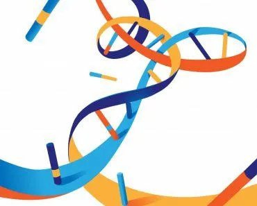
Chromatin is the complex of DNA and histone proteins that packages roughly 2 metres of human DNA into a cell nucleus only about 5 micrometres across. Its basic repeating unit is the nucleosome, about 147 base pairs of DNA wound around an octamer of histones. Chromatin exists in two main states: loose, transcriptionally active euchromatin and densely packed, mostly inactive heterochromatin. During eukaryotic cell division, it condenses further into the rod-shaped chromosomes visible under a microscope.
Cells are the basic functional units of life. The major component in a cell is the genetic material, commonly referred to collectively as the DNA. It contains hereditary information that is passed on from parent to offspring during cell division. The DNA is well organized in eukaryotes and is present within the nucleus. Chromatin plays a major role in wrapping up the genetic material and housing it within the cell nucleus, but what exactly is chromatin? What is it composed of? What are its types? What role does it play in cell division? How are chromatin, chromosomes and chromatids related? These are just a few of the questions we will be trying to answer in this article.
What Is Chromatin?
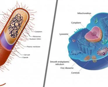
The DNA of prokaryotic cells posses a minimal amount of information, so it is simply distributed in a circular form over the cytoplasm. However, the DNA of eukaryotes contains millions of pieces of hereditary information. Therefore, it’s important to organize them properly in order to fit into the nucleus. Chromatin is a way to organize the genetic information to form the blueprint of life. It helps to pack the DNA into a small volume, so that it resides within the nucleus, with all the genetic information contained safely. It prevents the DNA from becoming tangled and plays a major role in reinforcing the DNA during cell division by regulating gene expression, facilitating DNA replication and preventing damage.
What Is Chromatin Composed Of?
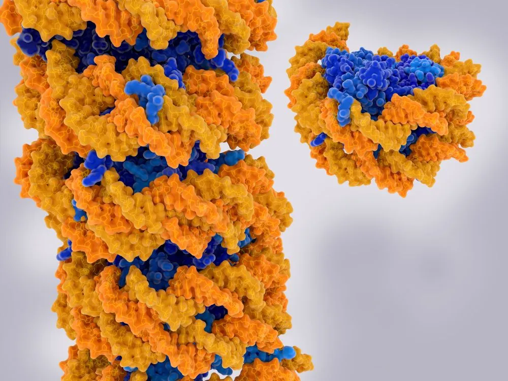
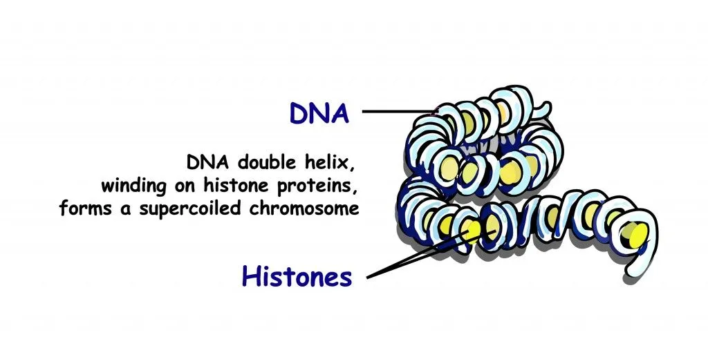
Chromatin is composed of two main parts, namely the DNA and the binding protein histone. Histone is an octomer composed of four sub-units that repeat twice. Histone acts as an anchor around which DNA is coiled. There are about 147 base pairs of DNA that wrap around the histones, forming nucleosomes. These nucleosomes are interconnected by sections of linker DNA. Histone can be classified into two types:
- Core histone
- Linker histone
H2A, H2B, H3 and H4 are the core histones. H1 is the linker histone that controls the entry and exit of a DNA strand on nucleosomes. Chemically, chromatin is composed of 30-40% DNA, 1-10% RNA and 50-60% proteins. This composition varies from one organism to another, in different tissues of the same species and in every stage of the cell cycle. This leads to the formation of more and less condensed regions within the structure.
Types Of Chromatin
Chromatin can be broadly classified into two types, based on its condensation:
Hetero-chromatin
Hetero-chromatin is a stable yet dynamic structure that varies from one cell to another. It is very tightly packed and has a highly condensed form. Being a repressive structure, it is inhibitory to the expression of genes within it. Higher order structures are formed by repetitive folding, which in turn increases the negative super-coiling of DNA. Within the hetero-chromatin, there are DNA structures called barriers that allow the expression of nucleosomes. Accessing the DNA is quite tedious in this form.
Eu-chromatin
Eu-chromatin consists of structures that are loosely packed. Modification to the histone tails allows them to be more open. This enables the easy access of DNA within these structures. It’s main function is the initiation of transcription. Eu-chromatin is actively involved in the transcription of DNA into mRNA. It also allows the recruitment of RNA polymerase complexes and gene regulatory proteins. The productivity of a cell is directly proportional to the amount of Eu-chromatin present within the cell.
Both these forms play their respective roles in gene transcription. Also, under necessary conditions, Eu-chromatin can be transformed into Hetero-chromatin. The cell cycle and other such processes use this ability to regulate the transcription of different genes.
Role Of Chromatin In Cell Division
Cell division is a process in which DNA produces multiple copies of itself, which can then be passed on to the next generation. During cell division, chromatin forms a more complex structure called a chromosome. These structures are visible only during mitotic cell division.
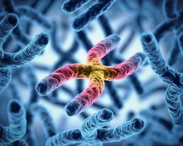
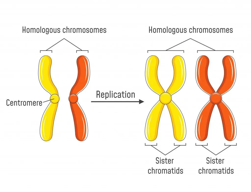
This visible condensation happens during prophase, but the doubling that creates the X-shape occurs earlier, during the synthesis (S) phase, when the DNA is replicated. That replication leaves each chromosome made of two identical copies, called sister chromatids, that stay attached to each other at the centromere until they are pulled apart later in division. To transcribe DNA into mRNA, or for the production of proteins, access to the DNA is essential. Since it is tightly wrapped around the histone, chromatin remodeling can be done to gain access to the desired segment of DNA where the gene is present.
In many events of cellular life, secondary structure of DNA is very important. Processes such as replication, transcription and regulation of expression of genes depend on the local changes in the DNA structure.
Recombination and specific mutations occur as a result of special changes in DNA. Chromatin controls all the above mentioned processes and is also responsible for hereditary changes.
What Is The Relationship Between Chromatin And Chromosomes?
This is the question most people actually arrive with, so let us answer it plainly: chromatin and a chromosome are the same material in two different states of packing. Chromatin is the everyday, working form of DNA and its histone proteins inside the nucleus. A chromosome is what that very same chromatin becomes when the cell condenses it down for division. Nothing is added or swapped out. The DNA, the histones and the nucleosomes are identical in both; only how tightly they are coiled changes.

For most of a cell's life (the long stretch called interphase) the chromatin sits relatively loose and thread-like, spread through the nucleus so the cell can read its genes, copy its DNA and repair damage. When the cell prepares to divide, that thin chromatin is wound up step by step (DNA around histones into nucleosomes, nucleosomes into a fiber, and that fiber into ever-tighter coils) until it forms the short, rod-shaped chromosomes you can finally see under a light microscope. The degree of squeezing is striking: in a metaphase chromosome the DNA has been condensed roughly 10,000-fold relative to its stretched-out length, which is how about 2 metres of DNA fits inside a nucleus only a few micrometres across.
So a useful rule of thumb: if the genetic material is unwound and being actively used, biologists call it chromatin; if it is condensed and ready to be parcelled out to daughter cells, they call it a chromosome. Same DNA, different job.
Chromatin Vs. Chromatid Vs. Chromosome: What Is The Difference?
A third word, chromatid, often joins the confusion. Here is how the three terms line up, moving from the least packed form to the most condensed and tracking them across the cell cycle:
- Chromatin is the loosely organized complex of DNA and histone proteins, the form the genome takes while the cell is going about its normal business.
- Chromosome is the highly condensed form of that same chromatin, compact enough to be moved cleanly during cell division.
- Chromatid is one of the two identical copies of a chromosome produced when the DNA is duplicated before division. The two are called sister chromatids, and they stay joined at a pinched region called the centromere.

The timing is the key that ties it together. During the synthesis (S) phase, the cell copies all of its DNA, so each chromosome that began as a single chromatid now consists of two identical sister chromatids held together at the centromere. That familiar X-shape you picture for a chromosome is actually a replicated chromosome: two sister chromatids waiting to be pulled apart. When division reaches anaphase, the centromere releases and the sister chromatids are drawn to opposite ends of the cell, where each becomes a chromosome in its own right inside a new daughter cell. Through all of this the underlying substance never stops being chromatin; chromatid and chromosome are simply names for what that chromatin looks like at particular moments. A human body cell, for the record, carries 46 chromosomes (23 pairs) packaged from this same chromatin.
References (click to expand)
- Chromatin.
- Swift, H. (1974, January 1). The Organization of Genetic Material in Eukaryotes: Progress and Prospects. Cold Spring Harbor Symposia on Quantitative Biology. Cold Spring Harbor Laboratory.
- Chromosomes and Chromatin. The Cell: A Molecular Approach (Cooper). NCBI Bookshelf.
- Chromosomal DNA and Its Packaging in the Chromatin Fiber. Molecular Biology of the Cell. NCBI Bookshelf.
- Chromatid. National Human Genome Research Institute (genome.gov).